
The Nervous System (NS) consists of the brain, the spinal cord and the nerves. It is a device for fast control of activities in a co-ordinated manner. It enables an individual to:-
a) Respond in a co-ordinated manner to environmental changes
b) Control various movements
c) Prolong life by protecting the body against harmful stimuli
There are about 1011 neurons in our NS. The NS originates from ectoderm. Anatomically NS can be classified as:-
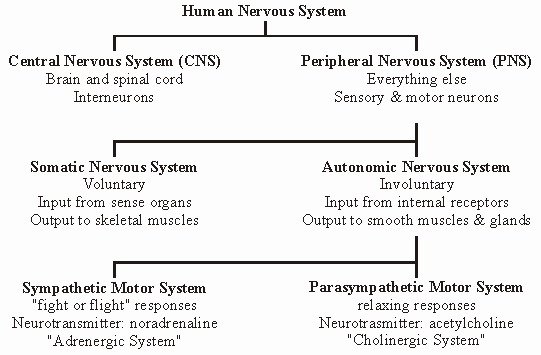
The Somatic NS consists of 12 pairs of cranial nerves and 31 pairs of spinal nerves.
THE NEURON
“Neuron (nerve cell) is the structural and functional unit of the NS”. Only the microglial cells are derived from mesoderm whereas, other neurons are ectodermal in origin.
MORPHOLOGY: A neuron consists of:-
F Cyton (cell body or soma or perikaryon).
F Dendron, which is a short process arising from cyton.
F Axon, which is a long process arising from cyton.
The dendrons give rise to dendrites. It is a
rule that, “dendrites convey impulses towards the soma and
axon away fro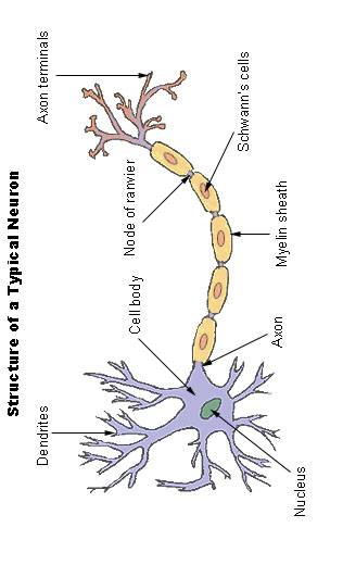 m
soma”.
m
soma”.
NEURONE: It is the component unit of a nervous pathway formed by a single neuron with its processes. It is also called as a nerve unit.
The axon and dendrons differ in many ways like length, thickness, nature of division, numbers in each cell, structure, external appearance and nature of nerve conduction. Like all other somatic cells, the neuron contains cytoplasm, nucleus, nucleolus, mitochondria and Golgi apparatus.
v Neurons cannot divide due to the absence of the centrosomes.
NEUROFIBRILS: An important fibrillary structure present in soma as well as dendron, with function unknown yet.
NISSL’S BODIES OR GRANULES: Small, angular, basic-staining granules within the neurone. They are absent in the axon hillock and this is made use of in distinguishing axon from dendron. Chemically, these granules are RNA and polysomes with organic Fe.
NEURONE – CLASSIFICATION
1. FUNCTIONAL
a) Motor/Efferent: They carry motor or volitional impulses from brain and spine to effector organs. These are further divided in to upper and lower motor neurons.
b) Sensory/Afferent: They carry impulses from periphery to brain and spine.
c) Intercalated/Internuncial: They form synaptic connection between afferent and efferent neurons.
2. LENGTH OF AXON
a) Golgi type – I: They have long axis cylinder that are able to connect with distant organs and cells. They have more cytoplasm.
b) Golgi type – II: Very small and short axons. Found in layer IV of cerebral cortex. Also present in spinal cord as intercalated neurons.
3. ACCORDING TO POLE
a) Unipolar: Both axon and dendron arise from a dichotomously splitting pole. Not usually found in adults.
b) Bipolar: Fusiform in shape. Two poles giving rise to either axon or dendron.
c)
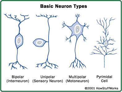 Multipolar:
Have multiple poles with processes, of which one is axon and all others are
dendrons.
Multipolar:
Have multiple poles with processes, of which one is axon and all others are
dendrons.
4. SHAPE
a) Triangular: They give out processes at the base. Found in cerebral cortex. Also known as pyramidal cells, giant pyramids and Betz cells.
b) Purkinje cells: Flask-shaped cells found in cerebellum.
c) Ganglion cells: Present in optic nerve.
5. AXON TERMINAL
a) Simple: Axon branches like a tree to form naked terminals.
b) Covered: These axons are meant to receive and respond to a stimulus. Thus, they end with receptors.
NEUROGLIAL CELLS: In addition to neurons that transmit nerve impulses, we also have supporting cells that have no physiological importance in nerve transmission. These cells are of various types and are present surrounding the neurons. They are called glial cells. Degenerated neurons are replaced by glial cells. The types of glial cells are:
Ø Astroglials: Provides support to neurons.
Ø Oligodendroglials: Provides protection to neurons.
Ø Microglials: Provides nutrition to neurons.
These glial cells also help in the formation of myelin sheath and phagocytosis.
THE NERVE OR NERVE FIBRE
 Nerves are axonal processes of neurons together with their covering sheaths.
They are white, glistening thread-like structures that are widely distributed.
The axons are surrounded by cell membrane of the Schwann cell.
Excitation and Conduction are the 2 important properties of nerves.
Nerves are axonal processes of neurons together with their covering sheaths.
They are white, glistening thread-like structures that are widely distributed.
The axons are surrounded by cell membrane of the Schwann cell.
Excitation and Conduction are the 2 important properties of nerves.
NERVE – CLASSIFICATION
1. STRUCTURAL
a) Myelinated/Medullated: These are the nerves that are covered by myelin sheath. Myelin sheath is a highly refractile, whitish, double contoured sheath, which closely invests large axons and dendrons. Both PNS and CNS have these types. But, the smallest axons in PNS do not have this sheath.
b) Non-myelinated/Non-medullated: They are not covered by myelin sheath. All post-ganglionic autonomic fibres, grey matter fibres and C-fibres have these.
2. FUNCTIONAL
a) Thickness and rate of transmission (table next page).
b) Type of impulse conducted: Motor and sensory nerves.
|
NERVE (alphabetical) |
TYPE (numerical) |
DIAMETER, µm |
VELOCITY OF TRANSMISSION, m/s |
FUNCTION |
|
A |
- |
2 – 20 |
12 – 120 |
Somatic, Motor and sensory |
|
Aα |
IA |
12 – 20 |
70 – 120 |
α-motor, Golgi tendon |
|
Aβ |
II |
5 – 12 |
30 – 70 |
Touch, Pressure |
|
Aα |
IB |
12 – 20 |
70 - 120 |
α-motor, Golgi tendon |
|
Aγ |
- |
3 – 6 |
15 – 35 |
Motor neurons to muscle spindles |
|
Aδ |
III |
2 – 5 |
12 – 30 |
Pain, Cold, touch |
|
B |
- |
1 – 3 |
3 – 15 |
Medullated autonomic preganglionic |
|
C |
IV |
0.2 – 1.5 |
0.4 – 2.3 |
Unmyelinated autonomic and somatic, Pain |
|
C |
IV |
0.2 – 1.5 |
0.4 – 2.3 |
Post-ganglionic sympathetic |
3. DEVELOPMENTAL
a) Somatic: An aggregation of parallel nerve fibres situated peripherally to CNS. They are divided in to afferent and efferent.
b) Visceral or autonomous: They supply the viscera, blood vessels and muscles. They are also divided in to afferent and efferent.
4. SOURCE OF ORIGIN
a) Cranial: May be entirely motor or sensory or mixed. The mixed nerves are attached to the same point on the surface of a brain stem.
b) Spinal: Always mixed type. Attached to surface of spinal cord by motor and sensory roots that have 2 different points of attachment.
5. CHEMICAL
a) Adrenergic: Synthesize and releases adrenaline (E) and nor-adrenaline (NE).
b) Cholinergic: Synthesize and releases acetyl choline (Ach).
NERVE NUCLEUS: Any localized collection of cells within CNS that have the same function. Eg: sensory and motor nuclei.
NERVE GANGLION: Any localized collection of cells within PNS that have the same function. They act as cell station. The cells of sympathetic and p-sympathetic ganglia are motor, whereas, those of cranio-spinal are sensory.
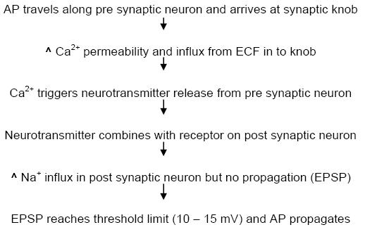
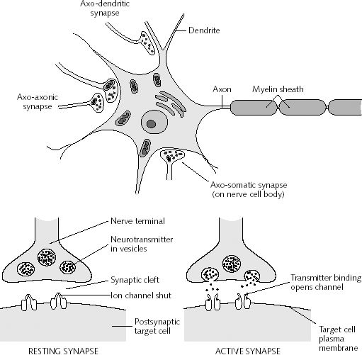
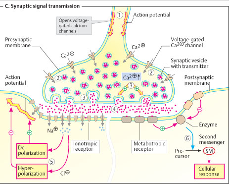
SYNAPSE
It is the junction where two neurons meet and permits impulse transmission.
· The axon has a plexus of fine branches ending in synaptic knob or end-feet (0.5 µ).
· Between the synaptic knob and cytoplasm, lies a membrane known as synaptolemma.
· The distance between pre-synaptic and post-synaptic neurons is about 20 – 40 nm.
· Due to this, the velocity of nerve impulse is retarded at synapse. This is known as synaptic delay or end-plate delay.
· A synapse may be incapable of transmitting an impulse. This is known as synaptic block.
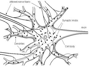
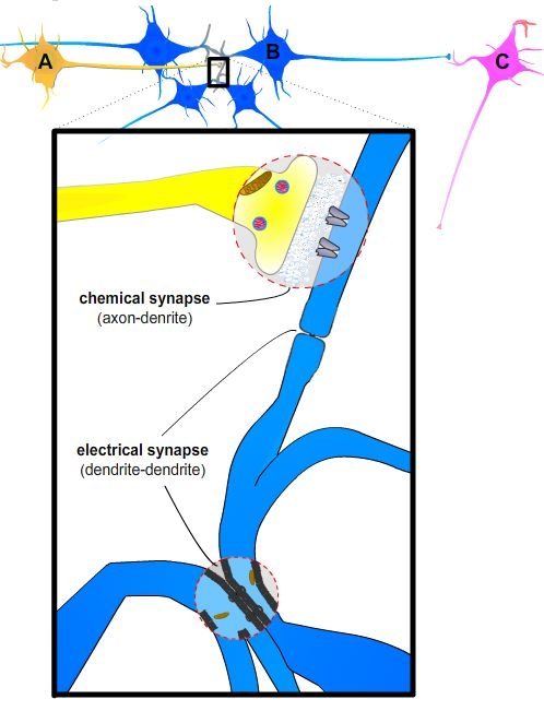
v LAW OF DYNAMIC POLARITY or SHERRINGTON’S LAW: It states that, “a synapse always passes an impulse from axon of one neuron to the dendron or soma of the next neuron and not in reverse direction”.
NERVE IMPULSE TRANSMISSION – IMPORTANT DEFINITIONS
1. NERVE IMPULSE: The physico-chemical changes brought about in response to a stimulus which propagates through the whole neurone.
2. BIOELECTRIC POTENTIAL: Electrical potential existing between inside and outside of a cell membrane of a living system. They are especially present in and function in nerves and muscles.
3. DIFFUSION POTENTIAL (or MEMBRANE POTENTIAL or JUNCTION POTENTIAL): Potential arising across a permeable membrane or at the junction between 2 solutions having different amounts of the same electrolyte.
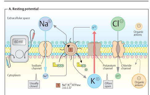
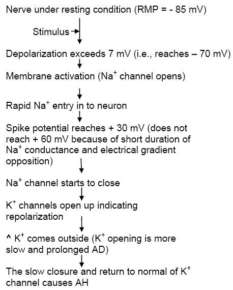
4. RESTING MEMBRANE POTENTIAL: It arises due to polarized condition of the plasma membrane separating the inside (-ve) and outside (+ve). Usually denoted with –ve sign. Value: - 50 to - 100 mV (or meV).
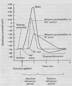
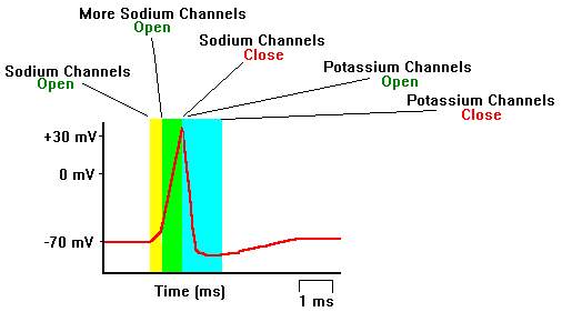
5. ACTION POTENTIAL: Reversal of RMP for a very short period on applying a stimulus. Value: + 30 to + 60 mV. It consists of 4 stages, namely:
a) Depolarization: Usually RMP = - 85 mV. When an impulse comes, polarization gets reversed, i.e., inside (+ve) and outside (-ve). This is known as depolarization. It rises sharply when RMP attains – 70 mV. It is also known as spike potential.
b) Repolarization: The restoration of normal RMP within a short time (1/2 of a msec). It starts fast but becomes slower and takes a longer period to reach the normal state.
c) Negative after potential: Also known as After depolarization (AD). The slower and longer period taken to reach normal RMP (2 – 15 msec).
d) Positive after potential: Also known as After hyperpolarization (AH). Even after AD, the membrane potential becomes even more – ve than the normal (- 85 mV) and remains –ve for about 50 msec or longer.
6.
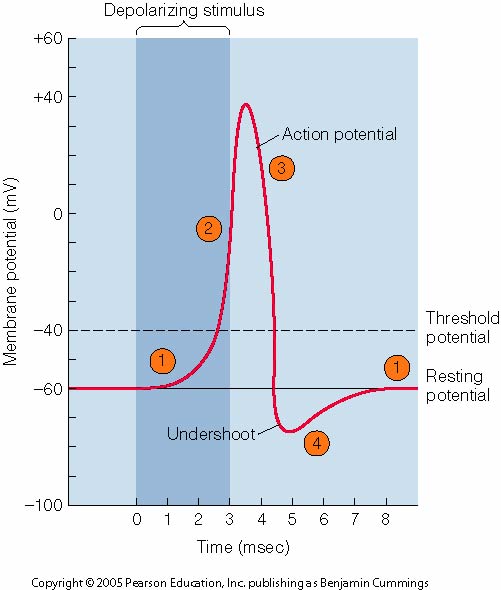 FIRING
LEVEL: It is the threshold value
needed to cause depolarization, i.e., - 85 to – 70 mV. Only on reaching this
level, AP will be propagated.
FIRING
LEVEL: It is the threshold value
needed to cause depolarization, i.e., - 85 to – 70 mV. Only on reaching this
level, AP will be propagated.
7. NERNST EQUATION: This is used to measure membrane potential.
E = [(u – v)/(u + v)] 2.303 RTF log (C1/C2)
Where, u = mobility of monovalent cation
v = mobility of monovalent anion
R = gas constant (Cal mole-1 oC-1)
T = absolute temperature (K)
F = faraday (96464 C mole-1),
C1 and C2 = concentration of electrolytes
IONIC BASIS OF RMP & AP
|
ION |
CONC, mEq L-1 H2O |
EQUILIBRIUM POTENTIAL, meV |
|
|
IN |
OUT |
||
|
Na+ |
15 |
150 |
+60 |
|
K+ |
150 |
5.5 |
-90 |
|
Cl- |
9 |
125 |
-70 |
|
A- |
65 |
0 |
- |
[A- is impermeable anions]
· From the table, it is clear that there is diffusion and concentration gradient setup across the cell membrane.
· For each ion tending to diffuse along its conc. gradient, a MP will be set up at rest, resulting in a equilibrium potential, which results in setting up of an electrical gradient.
· According to Nernst eqn., the equilibrium potentials were calculated for these permeable ions and given in the table.
· In fact K+ has observed equilibrium potential of – 70 mV, instead of – 90 mV, which is due to the fact that, a part of this high [K+] in, which is not taking part in diffusion is freely permeable that goes inside the cell.
· Na+ and K+ are actively transported out and in, respectively, by Na+/K+-ATPase pump.
· K+ movement out of the cell is along conc. gradient but against electrical gradient.
· K+ has ^ permeability [50 times more than Na+], so that, at equilibrium there is more K+ outside.
· For every 2 K+ pumped out, 3 Na+ is pumped in.
· The 3 factors for RMP are: - a) K+ permeability; b) A- impermeability; c) Na+/K+-ATPase pump.
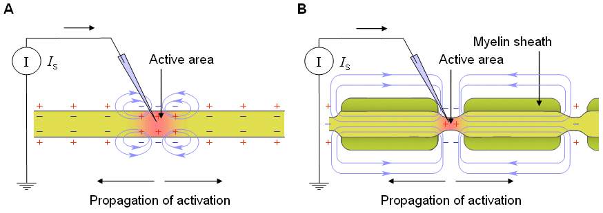
A) IMPULSE TRANSMISSION IN UNMYELINATED NERVES
[Ca2+ remains temporarily bound to Na+ permeable sites thereby inhibiting Na+ going inside during the resting state. Ca2+ gets removed from such sites on giving a stimulus]

B) IMPULSE TRANSMISSION IN MYELINATED NERVES
(SALTATORY PROPAGATION)
Ø The most excitable regions in the myelinated nerves are the nodes of Ranvier.
Ø It has 2000 – 12000 Na+ channels per µm2 compared to < 25 on myelin sheath surface.
Ø K+ channels are also located along side the Na+ channels.
Ø A current sink is set up in one node due to activation, causes depolarization of the next node which reaches firing level and thus, becomes activated.
Ø Thus, the impulse jumps or leaps from one node to another, because, myelin sheath acts as a good insulator.
Ø The impulse may also jump 2 – 3 nodes.
Velocity of impulse (ms-1) = diameter of fibre (µm) x 6
SIGNIFICANCE:
SYNAPTIC TRANSMISSION IN AUTONOMIC GANGLIA
SYNAPSE – TYPES: - a) Axo-somatic, b) Axo-axonic, c) Axo-dendritic (diagram next page)
Excitatory Post Synaptic Potential (EPSP): Depolarization caused by stimulation of sensory post synaptic neuron. Begins 0.5 msec, reaches peak after 1 – 1.5 msec and then declines exponentially with 4 msec. During this stage, the post synaptic neuron shows ^ sensitivity to other incoming impulses giving rise to EPSP.
Inhibitory Post Synaptic Potential (IPSP): Hyperpolarization of certain afferent neurons occurs 1 – 1.25 msec after impulse, reaches peak in 1.5 – 2 msec and declines exponentially within 3 msec. The excitability of post synaptic neuron is decreased, resulting in ^ K+ and Cl- permeability, but not to Na+.
The sequences of events that take place are:

SYNAPTIC TRANSMISSION IN CNS
This is more complex than that of autonomic ganglia, because:
a) Due to structural complexity (presence of intercalated neurons)
b) An afferent fibre ending in a synapse may either be excitatory or inhibitory
c) Several different chemical neurotransmitters involved
Here, the transmission is therefore of 2 types, namely: 1. Excitatory and 2. Inhibitory. Excitatory transmission in CNS synapse is same as that of autonomic ganglia.
Inhibitory transmission that involved no damage to the nerve occurs due to an ^ in RMP at the post synaptic neuron, i.e., hyperpolarization instead of depolarization. It is of 3 types: -
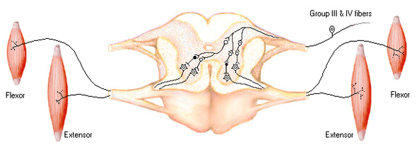
1. Direct (or) Post synaptic inhibition: Here, the intercalated neuron is present in between the afferent neuron and anterior horn cell (AHC).
a) The incoming AP via the intercalated neuron (called as Golgi bottle neuron) releases neurotransmitter
b) ^ Cl- influx (not Na+) in post synaptic neuron
c) ^ K+ efflux or closure of Na+/K+ channels produces slow IPSP
2. Pre synaptic inhibition: The intercalated neuron (called D-cell) ends on the knob of an excitatory neuron through axo-axonal synapse.
a) ^ Cl- influx in pre synaptic neuron
b) Decreased Ca2+ influx in pre synaptic neuron
c) Inhibition of neurotransmitter release
3. Recurrent (or) Renshaw cell inhibition: The intercalated neuron is called Renshaw cell. The spinal motor neuron gives off a branch, which connects with the Renshaw cell within the AHCs. This provides a negative feed back inhibition and helps to prevent excess discharge of impulse.
SYNAPSE – PROPERTIES
F Obeys the Law of dynamic polarity.
F Synaptic delay: A delay occurs for atleast 0.5 msec in a synapse because of the time required for the events taking place at the cleft.
F Susceptible to fatigue, lack of O2, anesthetics and some drugs.
F Excitability increases in acidic condition and decreases in alkaline condition.
F Several properties of reflex arc coincide with those of synapse.
NERVE FIBRE – PROPERTIES
1. IRRITABILITY or EXCITABILITY: It is the ability to respond to a stimulus. It can be modified depending on many factors such as:-
a) strength of stimulus; b) frequency of stimulus; c) duration of stimulus; d) direction of current; e) presence or absence of injury; f) effect of internal or external agents.
2. CONDUCTIVITY: A nerve impulse can very well travel in both directions. But, it is always one-way across a synapse. The propagation of action potential (AP) from the site of excitation away from it is called conduction. It is of 2 types:
a) Orthodromic: Impulse travel from soma to axon to dendron.
b) Antidromic: Impulse travel from axon to soma to dendron. Usually, this is of less physiologic significance and gets blocked in the first synapse and fades away.
3. REFRACTIVITY:
a) Absolute refractory period: During rising and much of the falling phase of spike potential, the neuron is refractile (not responding) to further stimulation with same intensity.
b) Relative refractory period: During rising and much of the falling phase of spike potential, the neuron responds to stimulation with a stronger stimulus.
4. SUMMATION: Two consecutive subliminal stimuli separated by ≤ 0.5 msec, gives rise to a response due to addition of the effects.
5. ADAPTATION or ACCOMODATION: Neurones are less excitable when stimulated by slowly rising current strength. It is due to continued depolarization that ^ ses K+ permeability and inactivation of Na+ pump.
6. INDEFATIGABILITY: On repeated stimulation of a NM junction, the muscle fails to respond. On connecting the same nerve to another muscle, the muscle responds.
7. ALL-OR-NONE LAW: The nerve fibre under physiologic conditions will give either a maximum response or no response at all. In a nerve fibre, once a part is stimulated with a threshold stimulus, it sets up an AP which traverses the whole length of the fibre.
REFLEX ACTION
Reflex action is the involuntary response resulting from stimulation of receptor organs. It is: -
1. Involuntary and does not need consciousness.
2. Inborn (except conditioned reflex which is acquired).
3. Involves only spine and brain stem and not cerebral cortex (except conditioned reflex).
4. Present in all members of a given species (except conditioned reflex).
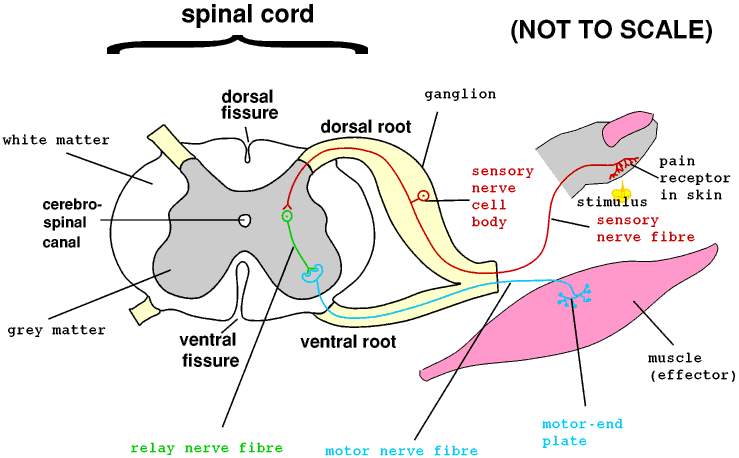
COMPONENTS: The components of reflex action are called as Reflex Arc. It consists of: -
1. An afferent limb (receptor organ + afferent neuron)
2. Reflex centre formed by synapse between afferent and efferent neurons situated in CNS.
3. Efferent limb (efferent neuron + effector organ/gland)
[NOTE: Afferent neuron is always dorsal and efferent neuron is always ventral]
CLASSIFICATION: Reflex can be classified in 10 different various ways. Each of the 10 groups has many sub-groups: -
1) Congenital or Acquired
2) Based on type of receptors
3) Based on purpose
4) Based on number of muscles responding
5) Based on effector organs
6) Based on parts of CNS involved
7) Based on character of response achieved
8) Clinical
9) Based on number of neuronal connections
Ø MONOSYNAPTIC (OR) 2 NEURONE REFLEX ARC: When the aff. and eff. neurons meet directly with only one synapse.
Ø DISYNAPTIC (OR) 3 NEURONE REFLEX ARC: When the aff. and eff. neurons are connected by an intercalated neuron.
Ø POLY (OR) MULTISYNAPTIC REFLEX ARC: Presence of more than one intercalated neuron between the aff. and eff. neurons.
10) Based on involvement of spinal segments
Ø CROSSED REFLEX ARC: The aff. and eff. neurons lie opposite to each other in spine.
Ø COMPLEX REFLEX ARC: They have innumerable connections through multisynaptic processes which involve many segments at various branches of spine and brain stem.
AXON REFLEX (ANTIDROMICITY)
It is a response of effector organ, produced by propagation of impulses from receptors along the aff. nerve and then through the branches of the axon of the same aff. nerve, to the effector. Simply, it is the conduction within one neuron which involves only a part of the neuron.
PROPERTIES: As seen previously, properties of reflex and synapse are almost the same, namely: -
a. Reflex impulse is always conducted in one-way manner (Bell-Magendie law).
b. Reflex delay is observed.
c. Susceptibility to fatigue.
d. Refractivity.
e. Summation.
f. Inhibition.
THE HUMAN BRAIN

The human brain is situated in the cranium (the bones which protect the brain), and it may be divided in to forebrain, midbrain and hindbrain. The hindbrain continues in to the spinal cord. These regions can be observed in the neural tube of a 6 wk embryo:
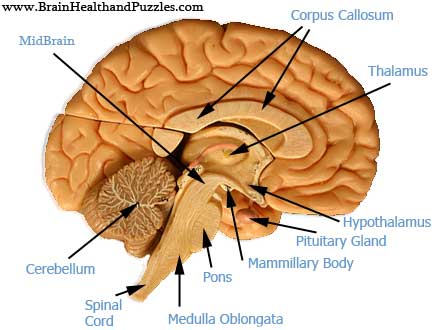
In most part of the brain, the grey matter containing the neurons is situated on the surface while the white matter made of fibres is located deep inside.
FOREBRAIN
F It consists of the cerebrum, the largest part of the brain, which is divided in two hemispheres (right and left).
F Corpus callosum is a curved thick bundle of nerve fibres which joins the 2 hemispheres at their base.
F 3 deep and wide fissures divide each cerebral hemisphere in to 4 lobes, viz., frontal, parietal, temporal and occipital. (see previous page)
F Cerebral cortex is the outer layer of the cerebrum and it is made of grey matter.
F Surface of each hemisphere shows many convolutions called gyri, which are separated by depressions called sulci. This arrangement ^ ses the surface area of cortex and accommodates more neurons.
FUNCTIONS OF FOREBRAIN
F Cerebral cortex is the highest centre for many sensations and activities like, learning, memory, cognition, control, emotions, etc.
F Somaesthetic area in the parietal lobe – perceptions like pain, touch and temperature.
F Motor area in frontal lobe – voluntary muscles.
F Premotor area in frontal lobe – involuntary muscles and ANS.
F Visual area in occipital lobe – vision.
F Auditory area in temporal lobe – hearing.
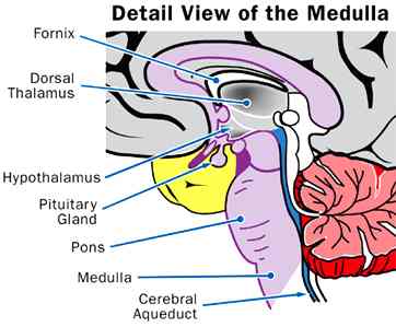
MIDBRAIN
It consists of many group of nerve nuclei scattered in white mater. Some of these are involved in controlling muscle tone and modify some motor activities initiated by cortex. Thalamus and hypothalamus are situated in the midbrain.
 FUNCTIONS
OF MIDBRAIN
FUNCTIONS
OF MIDBRAIN
F Thalamus forms conscious sensory centre, important relay station and also the reflex centre in man.
F Hypothalamus is associated with integrated control of both sympathetic and p-sympathetic systems, control of emotions, hunger, thirst, temperature, sleep and secretions of pituitary gland.
HINDBRAIN
It consists of the cerebellum located dorsally and the brain stem ventrally. The cerebellum is divided in to outer grey cerebellar cortex and inner medulla made of white matter. Cerebellar nuclei are also present in white matter. The brain stem consists of pons varoli and medulla oblongata, which extends to spinal cord.
FUNCTIONS OF HINDBRAIN
F Cerebellum contains centres for the maintenance of posture and equilibrium.
F They also modulate muscle tone of voluntary muscles, initiated by cerebral cortex.
F Brain stem has centres to control many vegetative activities, e.g. respiratory centre, vasomotor centre, salivary centre, etc.
F It also carries nerve tracts between the spinal cord and higher brain centres.
BRAIN – VENTRICLES
¨ Within the cerebral hemispheres, there are cavities called ventricles.
¨ Cerebrum contains 2 large cavities (lateral ventricle I and II) that lie lateral to the midline and a 3rd cavity (ventricle III) in the midline.
¨
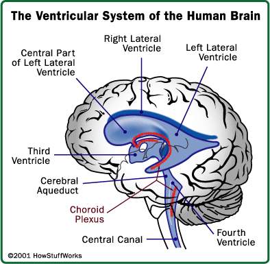 Each
lateral ventricle communicates with the III-v through the foramen of Monro.
Each
lateral ventricle communicates with the III-v through the foramen of Monro.
¨ Small cavities present in frontal, parietal and occipital lobes connect through small openings with III-v.
¨ From III-v, the cerebral aqueduct passes through the midbrain to open in to the IV-v of medulla oblongata that connects with spinal cord.
¨ The III-v is bound by thalamus and hypothalamus.
THE SPINE
§ Spinal cord is the 2nd part of the CNS, is the downward continuation of the medulla oblongata. It is cylindrical in shape.
§ It lies in the neural cavity of the vertebral column and reaches up to the level of the lower border of the first lumbar vertebra, where it ends in a conical extremity called the conus medullaris.
§ Beyond this, the vertebral column of the lumbar and sacral regions contains a bundle of nerves, called cauda equina or horse’s tail, rather than the spinal cord itself.
§ Spinal cord has 2 swellings namely cervical and lumbar enlargement.
§ The spinal cord is loosely surrounded by the 3 membranes or meninges which are continuous with those of the brain.
§ Below the conus medullaris, the pia mater is continued as a slender filament called the filum terminale.
§ Cross section of spine shows 2 fissures, namely, the anterior (mid-ventral, wide) and posterior fissures (mid-dorsal, narrow).
§ The outer portion of spine consists of white matter that contains the ascending (spine to brain) and descending (brain to spine regions) nerve tracts.
§ This white matter can be divided in to dorsal, lateral and ventral columns.
§ The grey matter is more centrally located and it contains the nerve soma and their synaptic processes. This region is in the form of letter “H” and can be divided in to dorsal, lateral and ventral grey horns.
§ A narrow central canal running through the mid of the grey mater communicates with the 4th ventricle of medulla oblongata.
SPINAL NERVES
§ 31 pairs of spinal nerves emerge from the sides of the cord.
§ Each nerve has an anterior (motor) and a posterior (sensory) root. The posterior root has ganglion.
§ At the end of dorsal grey horns, dorsal roots of spinal nerves are attached on each side. Similarly, the ventral nerves.
§ The dorsal and ventral roots join each other on each side of spine and lateral to the spine to form spinal nerve themselves.
FUNCTIONS OF SPINE
§ Have both motor and sensory functions.
§ Motor function may either be somatic or autonomic (cardiac and secretomotor).
§ Somatic consists of voluntary and involuntary motor functions.
§ Voluntary functions are carried out by anterior horn cells (AHC).
§ Sensory functions may be unconscious or conscious (exteroceptive, proprioceptive and enteroceptive).
CEREBROSPINAL FLUID (CSF)
CSF is the product of secretion of the choroid plexus. It is present in the sub-arachnoid space over the brain, brain ventricles and the spine. It is also present in the central canal of spine. The space between arachnoid membrane and piamater contains CSF.
FORMATION: It is formed in the choroid plexus of the lateral ventricles and also in the III and IV ventricles. The presence of mitochondria, granules and vesicles in the epithelial cells of the choroid plexus, indicate their capacity for active secretion. CSF cannot be an ultra-filtrate or dialysate of blood due to disagreement of observed physiologic parameters. Its rate of formation = 0.3 mL/min.
CIRCULATION
F From lateral ventricles it passes in to the III ventricle through the inter-ventricular foramen. (see brain ventricles diagram)
F From lateral ventricles it also passes in to the IV ventricle through aqueduct of Sylvius.
F Passes in to the subarachnoid space of brain and spine through foramen of Luschka and Magendi.
ABSORPTION: Mostly absorbed by the finger-like arachnoid villi present within the duramater in to the dural sinuses and spinal vein. A few amount is absorbed along the perineural spaces in to the cervical lymphatics and in to perivascular spaces.
FUNCTIONS:
Ø Acts as a fluid buffer and protects the vital neural substances.
Ø Regulates volume of cranial contents.
Ø Maintains proper ionic concentration for the CNS neurons.
Ø Acts as lymphatic system of CNS.
Ø Controls respiration.
Ø Acts as a medium for nutrient exchange as well as waste products.
THE BLOOD-BRAIN BARRIER (BBB)
This refers to the slow permeability or complete lack of permeability of a number of substances from the blood to the brain as well as to the CSF. It is due to: -
|
CSF constituents |
(mg/dL) |
|
Protein |
20 – 40 |
|
Glucose |
40 – 70 |
|
NaCl |
720 – 750 |
|
Cholesterol |
Nil or trace |
|
Urea |
10 – 30 |
|
Creatinine |
0.5 – 1.9 |
|
Uric acid |
0.5 – 2.5 |
|
Phosphates |
1.2 – 2.0 |
|
Na+ |
137 – 145 (mOsm/L) |
|
K+ |
2.7 – 3.9 |
|
Ca2+ |
2.1 – 3.0 |
|
Mg2+ |
2.0 – 2.5 |
|
Cl- |
116 – 122 |
|
HCO3- |
20 – 24 |
|
pH |
7.31 – 7.34 |
|
Osmolarity |
285 – 295 |
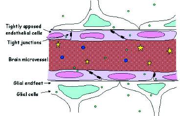
§ The endothelial cells of the brain capillaries have very tight junctions.
§ There are very few pores.
§ A dense protein matrix is also present between the endothelium and brain cells.
§ The brain capillaries are surrounded by a membrane made up of the end feet of astrocytes.
PERMEABILITY
a) Rapidly permeable: CO2, O2, H2O, L-Dopa, 5-OH tryptophan, lipid-soluble substances.
b) Slowly permeable: H+, HCO3-, Na+, K+, Mg2+, Ca2+, HPO42-, urea, and other water-soluble substances.
c) Glucose and amino acids pass through facilitated diffusion.
d) Impermeable: Large mol. wt. substances like bile salts and catecholamines.
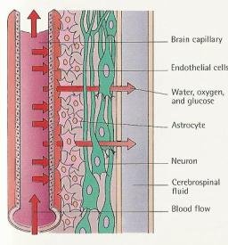
SIGNIFICANCE: It helps to maintain the internal milieu of neurons within the CNS.
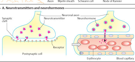
NEUROTRANSMITTERS (NT)
NTs are chemical substances synthesized in pre synaptic neuron and function to transmit impulses to the post synaptic neuron. A compound must satisfy the following criteria in order to be a NT: -
1. The pre synaptic neuron must have the substrates and enzymes necessary to synthesize the NT at the end feet.
2. The substance must be released by specific stimulation.
3. Micro application of the chemical to post synaptic neuron should mimic the effects of direct stimulation by pre synaptic neuron.
PUTATIVE NTs: These are those chemicals that are suspected to function as NT.
The nerve fibres that synthesize and release these NTs are broadly classified in to 2, namely: -
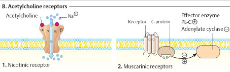
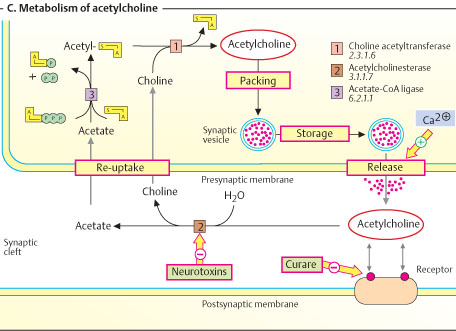
a. Cholinergic fibres: All pre ganglionic and post ganglionic fibres of p-sympathetic nervous system. They release Ach. They have of 2 types of receptors: Nicotinic cholinergic (nAchR) which is an ion-channel receptor and Muscarinic cholinergic (mAchR) which is a GPCR.
b. Adrenergic fibres: The post ganglionic sympathetic fibres. They release adrenaline or adrenaline-like substances. They act through GPCR.
NT – CHEMICAL CLASSIFICATION
|
CHEMICAL NATURE |
EXAMPLES |
|
Amine |
Dopamine, Serotonin, Ach, E, NE, etc. |
|
Amino acid |
A, D, E, G, Q, GABA |
|
Oligopeptide (neuro-active peptides) |
VIP, CCK, Motilin, CGRP |
|
Purine |
Adenosine, ATP |
|
Gas |
NO,CO |
[The following table is a simple representation about NTs – their name, precursors, location of neurons which produce them and functions.]
|
S.No. |
NAME |
PRECURSOR |
LOCATION OF NEURON |
FUNCTIONS |
|
A. AMINES |
||||
|
1 |
Norepinephrine/Noradrenaline |
F, Y |
HT, brain stem, sympathetic post ganglia |
Usually physiologic, especially affects cardiovascular system |
|
2 |
Epinephrine/Adrenaline |
F, Y |
Sympathetic post ganglia |
Usually metabolic. Some physiologic roles also |
|
3 |
Dopamine |
F, Y |
Basal ganglia, adrenal medulla |
Affects extra-pyramidal system behaviour and locomotor activity |
|
4 |
Dopa |
F, Y |
Basal ganglia, adrenal medulla |
Affects extra-pyramidal system behaviour and locomotor activity |
|
5 |
Serotonin |
W |
limbic system, cerebellum, spine |
Regulation of temperature, sensory perceptions, sleep and mood. It is converted to melatonin in pineal |
|
6 |
Acetyl choline |
Ace-CoA + choline |
Midbrain reticulum, corpus striatum |
Similar to p-sympathetic stimulation effects |
|
7 |
Histamine |
H |
Pituitary, various parts of CNS |
^ HCl secretion, modulates arousal, thirst, adenohypophyseal secretions, nociception, BP, allergies |
|
8 |
Agmatine |
R |
Brain |
Acts as endogenous ligand for imidazoline receptors |
|
B. AMINO ACIDS |
||||
|
9 |
G |
- |
Brain & spine |
Inhibitory in spine and brain stem, but, excitatory in brain |
|
10 |
E |
- |
Brain & spine |
Excitatory. Act through metabotropic and ionotropic receptors |
|
11 |
D |
- |
Brain, spine |
Not yet known well |
|
12 |
GABA |
E |
Especially CNS |
It composes of about 1/3rd of NTs in brain. Inhibitory effects on post synaptic motor neurons |
|
C. OLIGOPEPTIDES (NEURO-ACTIVE PEPTIDES) |
||||
|
13 |
Opioid (enkephalins, endorphins) |
Amino acids |
Brain, dorsal horn of spine |
Perception and integration of pain and emotions. Act through 3 types of receptors (µ, δ, κ) |
|
14 |
Substance P |
Amino acids |
HT, brain stem |
Slow pain pathway, axon reflex, myenteric reflex and neuro-endocrine regulation |
|
15 |
Vaso-active Intestinal Polypeptide (VIP) |
Amino acids |
Hippocampus, amygdala, HT |
Inhibitory in smooth muscles and endothelials. Excitatory in glandular cells. |
|
16 |
Gastrin, secretin, glucagon, GIP |
Amino acids |
Brain |
Miscellaneous |
|
17 |
Cholecystokinin (CCK) |
Amino acids |
Cerebral cortex, corpus striatum |
Neural pathway for satiety. Co-exists with dopamine in midbrain |
|
18 |
Neurotensin |
Amino acids |
HT and retina |
May have pituitary functions. Inhibits Purkinje cells. On injecting i.v., ^ ses glycemia and glucagon |
|
19 |
Neuropeptide Y |
Amino acids |
HT and NE-secreting neurons of ANS |
Through HT ^ ses appetite and food intake. Through ANS, augments vaso-constrictive effect of NE |
|
20 |
Calcitonin gene-related peptide (CGRP) |
Amino acids |
CNS, adrenal medulla, pars intermedia of pituitary, etc. |
α-type present in nerves. Very little effect on Ca2+ metabolism |
|
D. PURINES |
||||
|
21 |
Adenosine |
- |
CNS, PNS |
In CNS acts as depressant. In PNS, acts as vaso dilator |
|
22 |
ATP |
Adenosine |
Autonomic ganglia |
Rapid synaptic responses in ANS |
|
23 |
Caffeine & Theophylline |
- |
CNS, PNS |
Inhibitory to adenosine |
|
E. GASES |
||||
|
24 |
NO |
R |
Brain |
Also called as (EDRF:endothelium-derived relaxing factor). Easily crosses cell membrane. Activates GPCR-cGMP pathway. Also does cardiovascular regulation |
|
25 |
CO |
Heme to BV conversion |
Brain |
May activate GPCR-guanylate pathway |