HYPERSENSITIVITY
Introduction
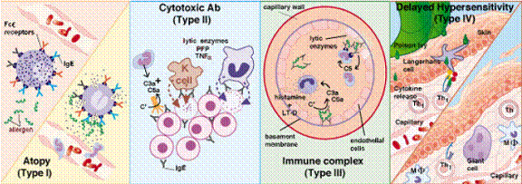
Clinical problems that have immune components arise when there is a loss of control of the immune response leading to tissue injury. These deleterious reactions are termed hypersensitivity, and the types are classified based on the mechanism by which the injury is caused . The types I, II and III are all mediated by antibody so effects are seen quickly; i.e., within a few minutes (allergic responses) to a couple of hours. Type IV is mediated by T cells particularly Th1 cells. Type IV injury is caused by accumulation of mononuclear cells at the site of reaction, so the peak response is not reached until between 24and 72 hours. It is important to remember that hypersensitivities are all secondary responses. The person must have become sensitized to the allergen (antigens that induce hypersensitivity reactions are usually termed allergens) by prior exposure. The greater extent and duration of the uncontrolled secondary response produces the injury. Almost any substance capable ofinducing an immune response is a potential allergen. Development of hypersensitivity to any particular allergen is due to a complex interaction of genetic susceptibility and exposure. (Allergic rhinitis due to ragweed pollen is common, but it is not problem for those who live in areas where ragweeds do not grow.) The necessity for exposure is the basis of the most common treatment, removal of the offending antigen. If you are allergic to poison ivy, wear gloves when pulling it our of your fence row! If avoidance in not possible, it may be necessary to use immunosuppression, either temporarily or on a more prolonged basis. A growing number of diseases/syndromes are being shown to have some symptoms due to immune reactions.

ALLERGY (Atopy or Type – I Hypersensitivity)
Introduction
Clinically important pathologies resulting at least in part from activities of the immune system arise when there is a loss of control of the immune response leading to tissue injury. The types of injury are classified based on the mechanism by which the injury is caused. Allergies are among the most common clinical complaints, affecting from 20 - 25% of the population. There is a genetic component to development of allergies in that 50% of the children have allergies when both parents have allergies. Even if only one parent has allergies, 30% of his or her children will also have allergies. The more serious forms of allergic reactions, asthma and anaphylaxis, can be life-threatening. When there is a failure of the normal controls on immune responses or an immune response is unduly prolonged, the immune response itself may cause damage to host tissues. Immune system mediated tissue injuries are classified according to the mechanism by which the injury is caused. Type I injury is mediated by IgE which is bound by specific receptors on mast cells. a. Cross-linking the bound IgE with the allergen stimulates the mast cell to release vasoactive substances, primarily histamine in humans. b. Symptoms may be localized or systemic depending on how the allergen is introduced.
Asthma
Asthma affects about 5% of the population. Of those, about half have attacks brought on by known allergens. Many of the others are thought to be triggered by allergens that have not yet been identified.
Pathophysiology: Allergens reaching the lungstrigger mast cell degranulation. Histamine induces constriction of bronchioles (H1 receptors) and relaxation of capillaries (H2receptors). C. TreatmentTreatment is usually viainhaled -andrenergic bronchodilators for acute attacks. Epinephrine is the most common prophylactic drug.
Anaphylaxis
Anaphylaxisis the response to systemic mast cell degranulation.
Symptoms: Systemic vasodilation can produce hypovolemic shock and death. Other organs strongly affected include the upper respiratory tract, skin, and GI tract.
Allergens: The allergen is usually from food, or is from a bite or sting. D. TreatmentTreatment includes aqueous epinephrine, tracheotomy (if laryngeal edema is severe), and antihistamines. Obviously avoidance is preferred.
Allergic Rhinitis
Allergic rhinitis is the most common immune mediated clinical entity. It may be seasonal or more-or-less continuous depending of the allergen.
Pathophysiology:
Allergen induced mast cell degranulation releases histamine and other vasoactive substances. The subsequent vasodilation produces the characteristic fluid exudate. Mast cells also release eosinophil chemotactic factor accounting for the presence of eosinophils in nasal exudate.C. TreatmentTreatment consists of avoidance, desensitization, and symptomatic relief.
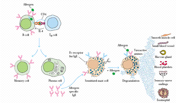
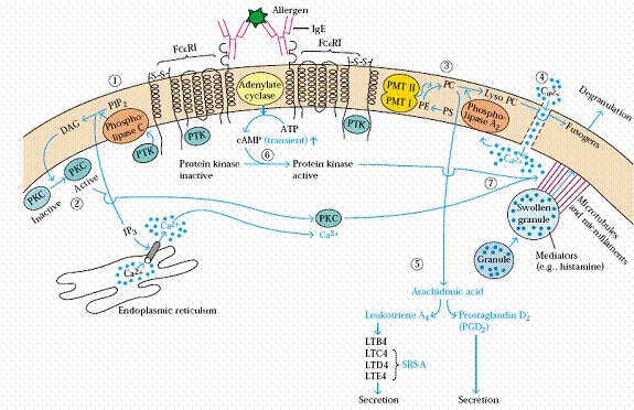
Mast cells and basophils have receptors specific for the Fc portion of IgE, so they are covered with IgE molecules picked up from plasma or tissue fluids. When the IgE on the mast cell or basophil is crosslinked by its antigen, the cell degranulates; i.e., releases all of its little bags of vasoactive substances. Histamine is released in the largest quantity and causes an immediate, localized inflammatory reaction. This produces the reddened eyes, edematous nasal mucosa, and rhinorrhea described above, but its effects wear off within an hour or so. However, leukotrienes C4, D2 & E4 (arachadonic acid metabolites as are prostaglandins) are also released by mast cell and basophil degranulation. Before they were isolated and characterized, these substances were known as slow reactive substance of anaphylaxis or SRS-A. They start to act about the time the effects of histamine wear off to produce a persistent inflammation. Heparin, with its anticlotting action, is also released during degranulation. Eosinophil chemotactic factor (ECF) released by mast cells attracts eosinophils to the area and accounts for the eosinophilia in the nasal discharge. Eosinophils are very important in host defenses against parasitic helminths. The fact of IgE mediated release of ECF from explains the evolutionary benefit of IgE.
Treatment
Treatment of allergic rhinitisis usually multifactorial. It includes environmental measures to reduce contact with the allergen(s), symptomatic relief with various drugs, and desensitization. Environmental measures to eliminate or avoid the allergen is often the simplest treatment. Obviously, this requires identification of the allergen(s) involved. It is often impractical to avoid pollens, although a well-timed vacation could be considered for very time limited pollens. Allergens at home can require special cleaning, sealing mattresses and/or pillows in plastic cases, removing carpets, etc. Allergens relating to the work place can be more troublesome. If the work routine cannot be modified sufficiently and hygiene measures are not efficacious, a change in occupation may be necessary. Symptomatic relief is provided by drug therapy. Antihistamines in either spray or oral form are the most commonly used drugs. Newer forms are available that produce much less drowsiness. Overuse of nasal sprays may lead to a rebound effect (rhinitis medicamentosa). Comolyn sodium in aerosols and less often in eye drops provide excellent relief when used prophylactically with few if any long-term side effects. It acts to stabilize the membranes of mast cells and basophils so that the vasoactive factors are never released. Nasal sprays containing a locally acting corticosteroid is used with some success in long-term prophylaxis. Corticosteroids are anti-inflammatory and cytotoxic to most leukocytes, but their exact mechanisms of action in relieving allergic rhinitis are unknown. It bypasses most of the side effects of systemic corcosteroid treatment. The slight burning and epistaxis associated with this spray does not appear to be dangerous. Long-term use should be monitored for for localized effects of cortisol; e.g., mucosal atrophy. Long term relief is provided by desensitization via intradermal injections of allergen mixtures. The goal is to induce the formation of IgG antibodies against the allergens which would then remove them from circulation before the IgE on the mast cells had a chance to see the allergen.
Type II Hypersensitivity : (Type II Tissue Injury)
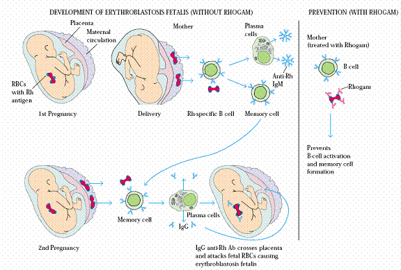
Cytotoxic antiself antibodies are responsible for type II injuries. The antibody may come from a transfusion, from the mother via the placenta erythroblastosis fetalis, or may be made by the host. If they cause tissue injury, it is, by definition, autoimmunity. When the antiself antibody bindsto its target tissue, it opsonizes the tissue for phagocytosis by macrophages and PMNs. When the phagocytes try to engulf the tissues, they release some lysosomal enzymes and reactive O2compounds. The injury to the surrounding tissue induces some inflammation. If the target antigen is on a cell surface, the antibody would be expected to disrupt cell function, and except for Graves' disease, would probably kill the cell. Complement would also be fixed if the antibody is IgM or IgG. This would exacerbate the injury by enhancing the inflammatory response and increasing the number of PMNs in the area. However, it is important to recognize that complement fixation is not required for tissue injury. Macrophages and PMNs will cause significant tissue injury in the complete absence of complement. If the target antigen is soluble, there may be formation of soluble immune complexes, so the tissue injury would result from type III mechanisms. This is the situation in systemic lupus erythematosisin which the target antigen is one or more proteins from the cell nucleus. If the autoantibody is being produced by the body rather than being passively acquired, it must be treated by immune suppression to prevent continued antibody production. Maintenance immunosuppression must usually be continued indefinitely.
Immune Complex Disease (Type –III Hypersensitivity):
The actual tissue injury is due to an intense, localized inflammatory reaction. The reaction is initiated by C3a and C5a produced during complement fixationinduced by immune complexes deposited in the tissue. Complement fixation is an absolute requirement for type III injury. C5b can sometimes bind to normal cells and provide the anchor for elaboration of the membrane attack complex which then lyses the cells. Tissue injury may activate the kinin system and produce platelet activating factor both of which intensify the inflammatory response. Much of the damage is caused by lysosomal enzymes and toxic oxygen products released into the tissues as PMNs try to engulf immune complexes. PMNs accumulate in the area in response to C5a and receptors expressed on endothelial cells in response to inflammatory signals. Immune complexes in the ratio of 2 antigens to 1IgG or IgA (monomeric) are often barely soluble. These complexes are often filtered out in capillary beds, particularly in the kidney and lungs, or may be driven into the vessel walls at places of especially turbulent flow. Larger, insoluble complexes are usually cleared quickly by phagocytic cells and cause little damage. This accounts for the typical kidney and lung damage produced by Type III injuries. Deposition at sites of turbulent flow with subsequent tissue injury at those sites is characteristic of polyarteritis nodosa.
Serum sickness:
It was originally seen following administration of antitoxin antiserum made in a different species usually a horse. This was once a common treatment for diphtheria and tetanus. Following administration of the horse serum, the patient made antibodies to the horse antigens. At the appropriate ratio, immune complexes were formed and deposited in the kidneys, lungs, and joints. Symptoms include fever, joint pain, dermatitis, lymphadenopathy, and in severe cases proteinuria and compromised pulmonary function. Mild forms may be seen following some vaccines or drug therapy.
Arthus reaction
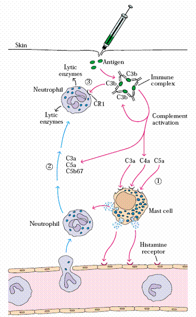
The model for immune complex diseases is the Arthus reaction, named after its discoverer in 1903. When antigen is injected intradermally into a hypersensitized animal, a localized cutaneous inflammatory reaction occurs that progresses to a hemorrhagic necrotic lesion. Histological examination of tissue biopsies of the site show an influx of PMNs. Staining with appropriate antibodies reveals immune complexes usually IgG-Ag and C3b. The injected antigen binds to antibody in the tissue fluids and forms Ag-Ab complexes that initiate complement fixation. C3a and C5a induce basophil degranulation and the inflammatory and PMN responses procede. The necrotic center often seen in an Arthus reaction is due to ischemia that results from platelet induced thrombi occluding the blood vessels at the site of the most intense reaction. Definitive diagnosis of immune complex disease requires determination of circulating immune complexes. However, reduced complement would be evidence in support of type III reactions occurring. Histological examination of the relevant tissue would show the immune complexes visualized by fluorescent labeled antibodies to human immunoglobulin in blobs. Treatment depends on the cause of the disease. If the antibody or antigen comes from an external source, one removes the source and provides symptomatic relief of the fever and kidney damage. If the source of the antigen is an infection, for example trypanosomiasis, treatment of disease to remove the source of the antigen would alleviate the problem. For an endogenous antigen i.e., antigen that arises from one's own tissues, it may not be feasible to eliminate the antigen. In these cases, immune suppression may be necessary.
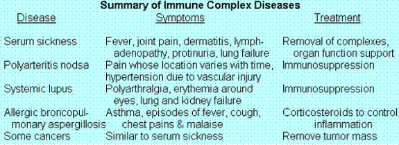
Diseases with an immunecomplex component are fairly common.
Systemic lupus erythematosus
It results from auto-antibodies against nuclear components (DNA, histones, etc.). These are normally sequestered inside the nucleus where they are hidden from the antibodies. They are released following tissue injury; e.g., a viral infection and are quickly bound by antibodies in the tissue fluids. The antigen-antibody complexes deposit in basement membranes of the golmeruli producing a lumpy-bumpy pattern and to a lesser extent lungs where it induces complement fixation, etc. Thus it is a combined type II and type III tissue injury. Patients usually die of kidney and/or lung failure. However, every organsystem may be involved. Polyarthralgia or arthritis is the most common symptom. It is four times more common in women than men and commonly strikes those of child bearing age. Treatment depends on the severity of the disease. In severe cases, corticosteroids, cyclophosphomide, and/or cyclosporin A may be used.
Delayed Hypersensitivity (Type –IV):
This is the only type of hypersensitivity mediated by T cells. Th1 cells respond preferentially to processed antigen on macrophage-lineage cells. Activated Th1 cells produce Il-2, GM-CSF, and IFN-all of which stimulate resting macrophages to become activated, "angry" macrophages with increased cytotoxic activity. In addition, Th1 cells secrete lymphokines (principally MIP-1and MIP-2) that attract macrophages and additional T cells to the site. They also secrete migration inhibitory factor (MIF) which prevents macrophages from leaving the area once they arrive. Activated macrophages exacerbate the problem by secreting IL-8 and IP-10 which are also chemotactic for mononuclear cells. So many mononuclear cells may accumulate in response to prolonged secretion of the chemotacticfactors that their pressure can block off arterioles. The tissue then dies from anoxia (lack of oxygen). Damage is exacerbated by release of hydrolytic enzymes, TNF-, prostaglandins, PFP, and toxic oxygen products from macrophages. Cytotoxic T cells may also make minor contributions to the tissue injury by releasing TNF-and additonal PFP. Since the injury is dependent on accumulation of T cells and macrophages, it usually takes 20 - 72 hours for symptoms to appear, but they may persist for a week or more. Individuals who are very sensitive to a particular substance may have symptoms as quickly as six hours.A delayed hypersensitivity reaction may be induced by recognition of "altered self" due to a drug or chemical binding to self protein; e.g., most penicillin reactions are due to this type of type IV reaction. They may also arise from persistent antigen the phagocytic system is unable to remove normally; e.g., schistosome eggs. In a few cases, Th cells become sensitized to self antigens and produce autoimmune tissue injury.
Diagnosis
Identification of the antigen is usually via patch-testing. Intradermal injection of small amounts of the suspected allergen is still used, particularly in testing for tuberculosis, but the patch test is easier and is less likely to produce necrosis. A good history will often provide needed clues. Definitive diagnosis may require histological examination of tissue samples. The particular symptoms developed depend on the tissue affected.
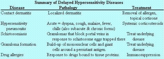
Delayed Hypersensitivity Diseases
Contact dermatitis
It is the most common and probably most pure form of delayed hypersensitivity. It is usually due to organic compounds attaching to proteins in the skin and acting as haptens. Heavy metals such an nickel a component of stainless steel may also act in this way. The combination is then taken up, by tissue Langerhans' cells, processed, and expressed on the cell surface in association with class II MHC proteins. Th1 cells are activated. The major symptoms are erythemic skin eruptions with swelling, vasculitis, and puritis. Rapidity of onset depends on the degree of sensitivity with a range of 6 hours to several days. Treatment consists of removal of the offending allergen and topical application of cortisone creams. The author knows of one case of a nearly fatal reaction following liberal application of sun tan lotion containing a mercury compound.
Mycobacteria such as those causing tuberculosis and leprosy are adapted to living inside cells, so they are somewhat resistant to killing by macrophages. Th1 cells continue to respond to the persistent antigens producing chemotactic factors on their own and stimulating macrophages to produce others. Continued production of the chemotactic factors induce granulomas to form around the bacteria. These serve to wall off the infection but may also compromise the function of the affected tissue. Treatment consists of alleviating the underlying disease.
Some drug allergies; e.g., penicillin, are due to their binding to serum or tissue proteins and acting as haptens to induce delayed hypersensitivity reactions similar to those for contact sensitivity. Immediate, IgE mediated, hypersensitivity reactions are more common. However, since the reaction is partly systemic, the results are much more serious than those seen in contact sensitivity reactions. Treatment may require temporary immunosuppression, usually with corticosteroids, until the drug-protein combination is cleared.
Mast Cell Degranulation – Type I hypersensitivity reactions
