At first glance, the petal of a flower or the skin on the back of a human hand may seem smooth and seamless, as if they were composed of a single, indistinct substance. In reality, however, many tiny individual units called cells make up these objects and almost all other components of plants and animals. The average human body contains over 75 trillion cells, but many life forms exist as single cells that perform all the functions necessary for independent existence. Most cells are far too small to be seen with the naked eye and require the use of high-power optical and electron microscopes for careful examination.
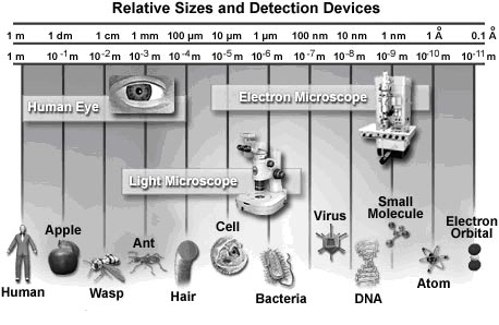
The relative scale of biological organisms as well as the useful range of several different detection devices is illustrated in Figure. The most basic intc sensor, the eye, was the only means humans had of visually observing the world around them for thousands of years. Though excellent for viewing a wide variety of objects, the power of the eye has its limits, anything smaller than the width of a single human hair being able to pass unnoticed by the organ. Therefore, when light microscopes of sufficient magnifying capability were developed in the late 1600s, a whole new world of tiny wonders was discovered. Electron microscopes, invented in the mid-twentieth century, made it possible to detect even tinier objects than light microscopes, including smaller molecules, viruses, and DNA. The detection power of most electron microscopes used today, however, stops just short of being able to visualize such incredibly small structures as the electron orbital systems of individual atoms. Atoms are considered the smallest units of an element that have the characteristics of that element, but cells are the smallest structural units of an organism capable of functioning independently.
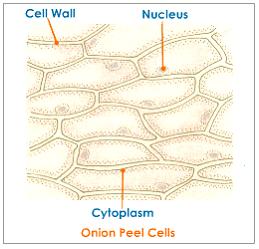
Yet, until the mid-seventeenth century, scientists were unaware that cells even existed. It wasn't until 1665 that biologist Robert Hooke observed through his microscope that plant tissues were divided into tiny compartments, which he termed "cellulae" or cells. It took another 175 years, however, before scientists began to understand the true importance of cells. In their studies of plant and animal cells during the early nineteenth century, German botanist Matthias Jakob Schleiden and German zoologist Theodor Schwann recognized the fundamental similarities between the two cell types. In 1839, they proposed that all living things are made up of cells, the theory that gave rise to modern biology.
Since that time, biologists have learned a great deal about the cell and its parts; what it is made of, how it functions, how it grows, and how it reproduces. The lingering question that is still being actively investigated is how cells evolved, i.e., how living cells originated from nonliving chemicals.
Numerous scientific disciplines—physics, geology, chemistry, and evolutionary biology—are being used to explore the question of cellular evolution. One theory speculates that substances vented into the air by volcanic eruptions were bombarded by lightning and ultraviolet radiation, producing larger, more stable molecules such as amino acids and nucleic acids. Rain carried these molecules to the Earth's surface where they formed a primordial soup of cellular building blocks.
A second theory proposes that cellular building blocks were formed in deep-water hydrothermal vents rather than in puddles or lakes on the Earth's surface. A third theory speculates that these key chemicals fell to earth on meteorites from outer space.
Given the basic building blocks and the right conditions, it would seem to be just a matter of time before cells begin to form. In the laboratory, lipid (fat) molecules have been observed joining together to produce spheres that are similar to a cell's plasma membrane. Over millions of years, perhaps it is inevitable that random collisions of lipid spheres with simple nucleic acids, such as RNA, would result in the first primitive cells capable of self-replication.
For all that has been learned about cells in over 300 years, hardly the least of which is the discovery of genetic inheritance and DNA, cell biology is still an exciting field of investigation. One recent addition is the study of how physical forces within the cell interact to form a stable biomechanical architecture. This is called "tensegrity" (a contraction of "tensional integrity"), a concept and word originally coined by Buckminster Fuller. The word refers to structures that are mechanically stable because stresses are distributed and balanced throughout the entire structure, not because the individual components have great strength.
In the realm of living cells, tensegrity is helping to explain how cells withstand physical stresses, how they are affected by the movements of organelles, and how a change in the cytoskeleton initiates biochemical reactions or even influences the action of genes. Some day, tensegrity may even explain the mechanical rules that caused molecules to assemble themselves into the first cells.
Animal Cells - Animal cells are typical of the eukaryotic cell type, enclosed by a plasma membrane and containing a membrane-bound nucleus and organelles.
Bacteria - One of the earliest prokaryotic cells to have evolved, bacteria have been around for at least 3.5 billion years and live in almost every imaginable environment.
Plant Cells - The basic plant cell has a similar construction to the animal cell, but does not have centrioles, lysosomes, cilia, or flagella. It does have additional structures, including a rigid cell wall, central vacuole, plasmodesmata, and chloroplasts.
Virus Structure - Viruses are not alive in the strict sense of the word, but reproduce and have an intimate, if parasitic, relationship with all living organisms.
Cells in Motion - In multicellular tissues, such as those found in animals and humans, individual cells employ a variety of locomotion mechanisms to maneuver through spaces in the extracellular matrix and over the surfaces of other cells. Examples are the rapid movement of cells in developing embryos, organ-to-organ spreading of malignant cancer cells, and the migration of neural axons to synaptic targets. Unlike single-celled swimming organisms, crawling cells in culture do not possess cilia or flagella, but tend to move by coordinated projection of the cytoplasm in repeating cycles of extension and retraction that deform the entire cell. The digital videos presented in this gallery investigate animal cell motility patterns in a wide variety of morphologically different specimens.
Fluorescence Microscopy of Cells in Culture - Serious attempts at the culture of whole tissues and isolated cells were first undertaken in the early 1900s as a technique for investigating the behavior of animal cells in an isolated and highly controlled environment. The term tissue culture arose because most of the early cells were derived from primary tissue explants, a technique that dominated the field for over 50 years. As established cell lines emerged, the application of well-defined normal and transformed cells in biomedical investigations has become an important staple in the development of cellular and molecular biology. This fluorescence intc gallery explores over 30 of the most common cell lines, labeled with a variety of fluorophores using both traditional staining methods as well as immunofluorescence techniques.
Observing Mitosis with Fluorescence Microscopy - Mitosis, a phenomenon observed in all higher eukaryotes, is the mechanism that allows the nuclei of cells to split and provide each daughter cell with a complete set of chromosomes during cellular division. This, coupled with cytokinesis (division of the cytoplasm), occurs in all multicellular plants and animals to permit growth of the organism. Digital imaging with fluorescence microscopy is becoming a powerful tool to assist scientists in understanding the complex process of mitosis on both a structural and functional level.
Cell Digestion and the Secretory Pathway - The primary sites of intracellular digestion are organelles known as the lysosomes, which are membrane-bounded compartments containing a variety of hydrolytic enzymes. Lysosomes maintain an internal acidic environment through the use of a hydrogen ion pump in the lysosomal membrane that drives ions from the cytoplasm into the lumenal space of the organelles. The high internal acidity is necessary for the enzymes contained in lysosomes to exhibit their optimum activity. Hence, if the integrity of a lysosomal membrane is compromised and the enzymatic contents are leaked into the cell, little damage is done due to the neutral pH of the cytoplasm. If numerous lysosomes rupture simultaneously, however, the cumulative action of their enzymes can result in autodigestion and the death of the cell.
Organisms may be broadly classified into two kinds:
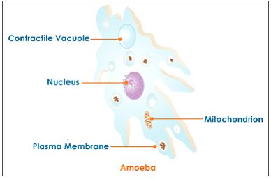
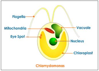
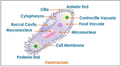
All living organisms, whether plants or animals are made up of microscopic units called cells. The cell occupies the same central position in biology as the atom in the physical sciences. All living beings, plants and animals, start their life with a single cell. Some organisms exist as a single cell and carry out the various metabolic life processes such as assimilation, respiration, reproduction, excretion, etc., that are essential for their survival. These are known as unicellular organisms.
Example:
Yeast, bacteria, chlamydomonas, amoeba
They are as unrelated to human beings as living things can be, but bacteria are essential to human life and life on planet Earth. Although they are notorious for their role in causing human diseases, from tooth decay to the Black Plague, there are beneficial species that are essential to good health.
For example, one species that lives symbiotically in the large intestine manufactures vitamin K, an essential blood clotting factor. Other species are beneficial indirectly. Bacteria give yogurt its tangy flavor and sourdough bread its sour taste. They make it possible for ruminant animals (cows, sheep, goats) to digest plant cellulose and for some plants, (soybean, peas, alfalfa) to convert nitrogen to a more usable form.
Bacteria are prokaryotes, lacking well-defined nuclei and membrane-bound organelles, and with chromosomes composed of a single closed DNA circle. They come in many shapes and sizes, from minute spheres, cylinders and spiral threads, to flagellated rods, and filamentous chains. They are found practically everywhere on Earth and live in some of the most unusual and seemingly inhospitable places.
Evidence shows that bacteria were in existence as long as 3.5 billion years ago, making them one of the oldest living organisms on the Earth. Even older than the bacteria are the archeans (also called archaebacteria) tiny prokaryotic organisms that live only in extreme environments: boiling water, super-salty pools, sulfur-spewing volcanic vents, acidic water, and deep in the Antarctic ice. Many scientists now believe that the archaea and bacteria developed separately from a common ancestor nearly four billion years ago. Millions of years later, the ancestors of today's eukaryotes split off from the archaea. Despite the superficial resemblance to bacteria, biochemically and genetically, the archea are as different from bacteria as bacteria are from humans.
In the late 1600s, Antoni van Leeuwenhoek became the first to study bacteria under the microscope. During the nineteenth century, the French scientist Louis Pasteur and the German physician Robert Koch demonstrated the role of bacteria as pathogens (causing disease). The twentieth century saw numerous advances in bacteriology, indicating their diversity, ancient lineage, and general importance. Most notably, a number of scientists around the world made contributions to the field of microbial ecology, showing that bacteria were essential to food webs and for the overall health of the Earth's ecosystems. The discovery that some bacteria produced compounds lethal to other bacteria led to the development of antibiotics, which revolutionized the field of medicine.
There are two different ways of grouping bacteria. They can be divided into three types based on their response to gaseous oxygen. Aerobic bacteria require oxygen for their health and existence and will die without it. Anerobic bacteria can't tolerate gaseous oxygen at all and die when exposed to it. Facultative aneraobes prefer oxygen, but can live without it.
The second way of grouping them is by how they obtain their energy. Bacteria that have to consume and break down complex organic compounds are heterotrophs. This includes species that are found in decaying material as well as those that utilize fermentation or respiration. Bacteria that create their own energy, fueled by light or through chemical reactions, are autotrophs.
· Capsule - Some species of bacteria have a third protective covering, a capsule made up of polysaccharides (complex carbohydrates). Capsules play a number of roles, but the most important are to keep the bacterium from drying out and to protect it from phagocytosis (engulfing) by larger microorganisms. The capsule is a major virulence factor in the major disease-causing bacteria, such as Escherichia coli and Streptococcus pneumoniae. Nonencapsulated mutants of these organisms are avirulent, i.e. they don't cause disease.
· Cell Envelope - The cell envelope is made up of two to three layers: the interior cytoplasmic membrane, the cell wall, and -- in some species of bacteria -- an outer capsule.
· Cell Wall - Each bacterium is enclosed by a rigid cell wall composed of peptidoglycan, a protein-sugar (polysaccharide) molecule. The wall gives the cell its shape and surrounds the cytoplasmic membrane, protecting it from the environment. It also helps to anchor appendages like the pili and flagella, which originate in the cytoplasm membrane and protrude through the wall to the outside. The strength of the wall is responsible for keeping the cell from bursting when there are large differences in osmotic pressure between the cytoplasm and the environment.
Cell wall composition varies widely amongst bacteria and is one of the most important factors in bacterial species analysis and differentiation. For example, a relatively thick, meshlike structure that makes it possible to distinguish two basic types of bacteria. A technique devised by Danish physician Hans Christian Gram in 1884, uses a staining and washing technique to differentiate between the two forms. When exposed to a gram stain, gram-positive bacteria retain the purple color of the stain because the structure of their cell walls traps the dye. In gram-negative bacteria, the cell wall is thin and releases the dye readily when washed with an alcohol or acetone solution.
· Cytoplasm - The cytoplasm, or protoplasm, of bacterial cells is where the functions for cell growth, metabolism, and replication are carried out. It is a gel-like matrix composed of water, enzymes, nutrients, wastes, and gases and contains cell structures such as ribosomes, a chromosome, and plasmids. The cell envelope encases the cytoplasm and all its components. Unlike the eukaryotic (true) cells, bacteria do not have a membrane enclosed nucleus. The chromosome, a single, continuous strand of DNA, is localized, but not contained, in a region of the cell called the nucleoid. All the other cellular components are scattered throughout the cytoplasm.
· Cytoplasmic Membrane - A layer of phospholipids and proteins, called the cytoplasmic membrane, encloses the interior of the bacterium, regulating the flow of materials in and out of the cell. This is a structural trait bacteria share with all other living cells; a barrier that allows them to selectively interact with their environment. Membranes are highly organized and asymmetric having two sides, each side with a different surface and different functions. Membranes are also dynamic, constantly adapting to different conditions.
One of those components, plasmids, are small, extrachromosomal genetic structures carried by many strains of bacteria. Like the chromosome, plasmids are made of a circular piece of DNA. Unlike the chromosome, they are not involved in reproduction. Only the chromosome has the genetic instructions for initiating and carrying out cell division, or binary fission, the primary means of reproduction in bacteria. Plasmids replicate independently of the chromosome and, while not essential for survival, appear to give bacteria a selective advantage.
Plasmids are passed on to other bacteria through two means. For most plasmid types, copies in the cytoplasm are passed on to daughter cells during binary fission. Other types of plasmids, however, form a tubelike structure at the surface called a pilus that passes copies of the plasmid to other bacteria during conjugation, a process by which bacteria exchange genetic information. Plasmids have been shown to be instrumental in the transmission of special properties, such as antibiotic drug resistance, resistance to heavy metals, and virulence factors necessary for infection of animal or plant hosts. The ability to insert specific genes into plasmids have made them extremely useful tools in the fields of molecular biology and genetics, specifically in the area of genetic engineering.
· Flagella - Flagella (singular, flagellum) are hairlike structures that provide a means of locomotion for those bacteria that have them. They can be found at either or both ends of a bacterium or all over its surface. The flagella beat in a propeller-like motion to help the bacterium move toward nutrients; away from toxic chemicals; or, in the case of the photosynthetic cyanobacteria; toward the light.
· Nucleoid - The nucleoid is a region of cytoplasm where the chromosomal DNA is located. It is not a membrane bound nucleus, but simply an area of the cytoplasm where the strands of DNA are found. Most bacteria have a single, circular chromosome that is responsible for replication, although a few species do have two or more. Smaller circular auxiliary DNA strands, called plasmids, are also found in the cytoplasm.
· Pili - Many species of bacteria have pili (singular, pilus), small hairlike projections emerging from the outside cell surface. These outgrowths assist the bacteria in attaching to other cells and surfaces, such as teeth, intestines, and rocks. Without pili, many disease-causing bacteria lose their ability to infect because they're unable to attach to host tissue. Specialized pili are used for conjugation, during which two bacteria exchange fragments of plasmid DNA.
· Ribosomes - Ribosomes are microscopic "factories" found in all cells, including bacteria. They translate the genetic code from the molecular language of nucleic acid to that of amino acids—the building blocks of proteins. Proteins are the molecules that perform all the functions of cells and living organisms. Bacterial ribosomes are similar to those of eukaryotes, but are smaller and have a slightly different composition and molecular structure. Bacterial ribosomes are never bound to other organelles as they sometimes are (bound to the endoplasmic reticulum) in eukaryotes, but are free-standing structures distributed throughout the cytoplasm. There are sufficient differences between bacterial ribosomes and eukaryotic ribosomes that some antibiotics will inhibit the functioning of bacterial ribosomes, but not a eukaryote's, thus killing bacteria but not the eukaryotic organisms they are infecting.
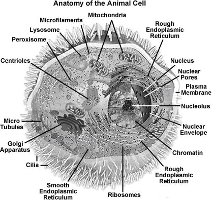
Animal cells are typical of the eukaryotic cell, enclosed by a plasma membrane and containing a membrane-bound nucleus and organelles. Unlike the eukaryotic cells of plants and fungi, animal cells do not have a cell wall. This feature was lost in the distant past by the single-celled organisms that gave rise to the kingdom Animalia. Most cells, both animal and plant, range in size between 1 and 100 micrometers and are thus visible only with the aid of a microscope.
The lack of a rigid cell wall allowed animals to develop a greater diversity of cell types, tissues, and organs. Specialized cells that formed nerves and muscles—tissues impossible for plants to evolve—gave these organisms mobility. The ability to move about by the use of specialized muscle tissues is a hallmark of the animal world, though a few animals, primarily sponges, do not possess differentiated tissues. Notably, protozoans locomote, but it is only via nonmuscular means, in effect, using cilia, flagella, and pseudopodia.
The animal kingdom is unique among eukaryotic organisms because most animal tissues are bound together in an extracellular matrix by a triple helix of protein known as collagen. Plant and fungal cells are bound together in tissues or aggregations by other molecules, such as pectin. The fact that no other organisms utilize collagen in this manner is one of the indications that all animals arose from a common unicellular ancestor. Bones, shells, spicules, and other hardened structures are formed when the collagen-containing extracellular matrix between animal cells becomes calcified.
Animals are a large and incredibly diverse group of organisms. Making up about three-quarters of the species on Earth, they run the gamut from corals and jellyfish to ants, whales, elephants, and, of course, humans. Being mobile has given animals, which are capable of sensing and responding to their environment, the flexibility to adopt many different modes of feeding, defense, and reproduction. Unlike plants, however, animals are unable to manufacture their own food, and therefore, are always directly or indirectly dependent on plant life.
Most animal cells are diploid, meaning that their chromosomes exist in homologous pairs. Different chromosomal ploidies are also, however, known to occasionally occur. The proliferation of animal cells occurs in a variety of ways. In instances of sexual reproduction, the cellular process of meiosis is first necessary so that haploid daughter cells, or gametes, can be produced. Two haploid cells then fuse to form a diploid zygote, which develops into a new organism as its cells divide and multiply.
Cells were discovered in 1665 by British scientist Robert Hooke who first observed them in his crude (by today's standards) seventeenth century optical microscope. In fact, Hooke coined the term "cell", in a biological context, when he described the microscopic structure of cork like a tiny, bare room or monk's cell. Illustrated in Figure 2 are a pair of fibroblast deer skin cells that have been labeled with fluorescent probes and photographed in the microscope to reveal their internal structure. The nuclei are stained with a red probe, while the Golgi apparatus and microfilament actin network are stained green and blue, respectively. The microscope has been a fundamental tool in the field of cell biology and is often used to observe living cells in culture. Use the links below to obtain more detailed information about the various components that are found in animal cells.
· Centrioles - Centrioles are self-replicating organelles made up of nine bundles of microtubules and are found only in animal cells. They appear to help in organizing cell division, but aren't essential to the process.
· Cilia and Flagella - For single-celled eukaryotes, cilia and flagella are essential for the locomotion of individual organisms. In multicellular organisms, cilia function to move fluid or materials past an immobile cell as well as moving a cell or group of cells.
· Endoplasmic Reticulum - The endoplasmic reticulum is a network of sacs that manufactures, processes, and transports chemical compounds for use inside and outside of the cell. It is connected to the double-layered nuclear envelope, providing a pipeline between the nucleus and the cytoplasm.
· Endosomes and Endocytosis - Endosomes are membrane-bound vesicles, formed via a complex family of processes collectively known as endocytosis, and found in the cytoplasm of virtually every animal cell. The basic mechanism of endocytosis is the reverse of what occurs during exocytosis or cellular secretion. It involves the invagination (folding inward) of a cell's plasma membrane to surround macromolecules or other matter diffusing through the extracellular fluid.
· Golgi Apparatus - The Golgi apparatus is the distribution and shipping department for the cell's chemical products. It modifies proteins and fats built in the endoplasmic reticulum and prepares them for export to the outside of the cell.
· Intermediate Filaments - Intermediate filaments are a very broad class of fibrous proteins that play an important role as both structural and functional elements of the cytoskeleton. Ranging in size from 8 to 12 nanometers, intermediate filaments function as tension-bearing elements to help maintain cell shape and rigidity.
· Lysosomes - The main function of these microbodies is digestion. Lysosomes break down cellular waste products and debris from outside the cell into simple compounds, which are transferred to the cytoplasm as new cell-building materials.
· Microfilaments - Microfilaments are solid rods made of globular proteins called actin. These filaments are primarily structural in function and are an important component of the cytoskeleton.
· Microtubules - These straight, hollow cylinders are found throughout the cytoplasm of all eukaryotic cells (prokaryotes don't have them) and carry out a variety of functions, ranging from transport to structural support.
· Mitochondria - Mitochondria are oblong shaped organelles that are found in the cytoplasm of every eukaryotic cell. In the animal cell, they are the main power generators, converting oxygen and nutrients into energy.
· Nucleus - The nucleus is a highly specialized organelle that serves as the information processing and administrative center of the cell. This organelle has two major functions: it stores the cell's hereditary material, or DNA, and it coordinates the cell's activities, which include growth, intermediary metabolism, protein synthesis, and reproduction (cell division).
· Peroxisomes - Microbodies are a diverse group of organelles that are found in the cytoplasm, roughly spherical and bound by a single membrane. There are several types of microbodies but peroxisomes are the most common.
· Plasma Membrane - All living cells have a plasma membrane that encloses their contents. In prokaryotes, the membrane is the inner layer of protection surrounded by a rigid cell wall. Eukaryotic animal cells have only the membrane to contain and protect their contents. These membranes also regulate the passage of molecules in and out of the cells.
· Ribosomes - All living cells contain ribosomes, tiny organelles composed of approximately 60 percent RNA and 40 percent protein. In eukaryotes, ribosomes are made of four strands of RNA. In prokaryotes, they consist of three strands of RNA.
In addition the optical and electron microscope, scientists are able to use a number of other techniques to probe the mysteries of the animal cell. Cells can be disassembled by chemical methods and their individual organelles and macromolecules isolated for study. The process of cell fractionation enables the scientist to prepare specific components, the mitochondria for example, in large quantities for investigations of their composition and functions. Using this approach, cell biologists have been able to assign various functions to specific locations within the cell. However, the era of fluorescent proteins has brought microscopy to the forefront of biology by enabling scientists to target living cells with highly localized probes for studies that don't interfere with the delicate balance of life processes.
Plants are unique among the eukaryotes, organisms whose cells have membrane-enclosed nuclei and organelles, because they can manufacture their own food. Chlorophyll, which gives plants their green color, enables them to use sunlight to convert water and carbon dioxide into sugars and carbohydrates, chemicals the cell uses for fuel.
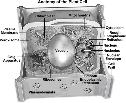
Like the fungi, another kingdom of eukaryotes, plant cells have retained the protective cell wall structure of their prokaryotic ancestors. The basic plant cell shares a similar construction motif with the typical eukaryote cell, but does not have centrioles, lysosomes, intermediate filaments, cilia, or flagella, as does the animal cell. Plant cells do, however, have a number of other specialized structures, including a rigid cell wall, central vacuole, plasmodesmata, and chloroplasts. Although plants (and their typical cells) are non-motile, some species produce gametes that do exhibit flagella and are, therefore, able to move about.
Plants can be broadly categorized into two basic types: vascular and nonvascular. Vascular plants are considered to be more advanced than nonvascular plants because they have evolved specialized tissues, namely xylem, which is involved in structural support and water conduction, and phloem, which functions in food conduction. Consequently, they also possess roots, stems, and leaves, representing a higher form of organization that is characteristically absent in plants lacking vascular tissues. The nonvascular plants, members of the division Bryophyta, are usually no more than an inch or two in height because they do not have adequate support, which is provided by vascular tissues to other plants, to grow bigger. They also are more dependent on the environment that surrounds them to maintain appropriate amounts of moisture and, therefore, tend to inhabit damp, shady areas.
It is estimated that there are at least 260,000 species of plants in the world today. They range in size and complexity from small, nonvascular mosses to giant sequoia trees, the largest living organisms, growing as tall as 330 feet (100 meters). Only a tiny percentage of those species are directly used by people for food, shelter, fiber, and medicine. Nonetheless, plants are the basis for the Earth's ecosystem and food web, and without them complex animal life forms (such as humans) could never have evolved. Indeed, all living organisms are dependent either directly or indirectly on the energy produced by photosynthesis, and the byproduct of this process, oxygen, is essential to animals. Plants also reduce the amount of carbon dioxide present in the atmosphere, hinder soil erosion, and influence water levels and quality.
Plants exhibit life cycles that involve alternating generations of diploid forms, which contain paired chromosome sets in their cell nuclei, and haploid forms, which only possess a single set. Generally these two forms of a plant are very dissimilar in appearance. In higher plants, the diploid generation, the members of which are known as sporophytes due to their ability to produce spores, is usually dominant and more recognizable than the haploid gametophyte generation. In Bryophytes, however, the gametophyte form is dominant and physiologically necessary to the sporophyte form.
Animals are required to consume protein in order to obtain nitrogen, but plants are able to utilize inorganic forms of the element and, therefore, do not need an outside source of protein. Plants do, however, usually require significant amounts of water, which is needed for the photosynthetic process, to maintain cell structure and facilitate growth, and as a means of bringing nutrients to plant cells. The amount of nutrients needed by plant species varies significantly, but nine elements are generally considered to be necessary in relatively large amounts. Termed macroelements, these nutrients include calcium, carbon, hydrogen, magnesium, nitrogen, oxygen, phosphorus, potassium, and sulfur. Seven microelements, which are required by plants in smaller quantities, have also been identified: boron, chlorine, copper, iron, manganese, molybdenum, and zinc.
· Cell Wall - Like their prokaryotic ancestors, plant cells have a rigid wall surrounding the plasma membrane. It is a far more complex structure, however, and serves a variety of functions, from protecting the cell to regulating the life cycle of the plant organism.
· Chloroplasts - The most important characteristic of plants is their ability to photosynthesize, in effect, to make their own food by converting light energy into chemical energy. This process is carried out in specialized organelles called chloroplasts.
· Endoplasmic Reticulum - The endoplasmic reticulum is a network of sacs that manufactures, processes, and transports chemical compounds for use inside and outside of the cell. It is connected to the double-layered nuclear envelope, providing a pipeline between the nucleus and the cytoplasm. In plants, the endoplasmic reticulum also connects between cells via the plasmodesmata.
· Golgi Apparatus - The Golgi apparatus is the distribution and shipping department for the cell's chemical products. It modifies proteins and fats built in the endoplasmic reticulum and prepares them for export as outside of the cell.
· Microfilaments - Microfilaments are solid rods made of globular proteins called actin. These filaments are primarily structural in function and are an important component of the cytoskeleton.
· Microtubules - These straight, hollow cylinders are found throughout the cytoplasm of all eukaryotic cells (prokaryotes don't have them) and carry out a variety of functions, ranging from transport to structural support.
· Mitochondria - Mitochondria are oblong shaped organelles found in the cytoplasm of all eukaryotic cells. In plant cells, they break down carbohydrate and sugar molecules to provide energy, particularly when light isn't available for the chloroplasts to produce energy.
· Nucleus - The nucleus is a highly specialized organelle that serves as the information processing and administrative center of the cell. This organelle has two major functions: it stores the cell's hereditary material, or DNA, and it coordinates the cell's activities, which include growth, intermediary metabolism, protein synthesis, and reproduction (cell division).
· Peroxisomes - Microbodies are a diverse group of organelles that are found in the cytoplasm, roughly spherical and bound by a single membrane. There are several types of microbodies but peroxisomes are the most common.
· Plasmodesmata - Plasmodesmata are small tubes that connect plant cells to each other, providing living bridges between cells.
· Plasma Membrane - All living cells have a plasma membrane that encloses their contents. In prokaryotes and plants, the membrane is the inner layer of protection surrounded by a rigid cell wall. These membranes also regulate the passage of molecules in and out of the cells.
· Ribosomes - All living cells contain ribosomes, tiny organelles composed of approximately 60 percent RNA and 40 percent protein. In eukaryotes, ribosomes are made of four strands of RNA. In prokaryotes, they consist of three strands of RNA.
· Vacuole - Each plant cell has a large, single vacuole that stores compounds, helps in plant growth, and plays an important structural role for the plant.
Leaf Tissue Organization - The plant body is divided into several organs: roots, stems, and leaves. The leaves are the primary photosynthetic organs of plants, serving as key sites where energy from light is converted into chemical energy. Similar to the other organs of a plant, a leaf is comprised of three basic tissue systems, including the dermal, vascular, and ground tissue systems. These three motifs are continuous throughout an entire plant, but their properties vary significantly based upon the organ type in which they are located. All three tissue systems are discussed in this section.
|
Animal cell |
Plant cell |
|
Usually smaller in size |
Usually larger in size |
|
Cell wall is absent. Cellulose in any form is absent |
Cell wall made up of cellulose is present |
|
Cytoplasm is denser, more granular and occupies most of the space in the cell |
Cytoplasm is pushed to the periphery and forms a thin lining against the cell wall. The central space in the cell may be occupied by a large, single vacuole |
|
Vacuoles absent. If present, they are small, temporary and concerned with excretion or secretion |
Vacuoles are large and prominent. May be one or more |
|
Plastids are absent |
Plastids are usually present |
|
Centrosome is present |
Centrosome is absent. Instead two small clear areas called polar caps are present |
|
Prominent and highly complex Golgi bodies present near nucleus |
Contain several sub units of Golgi apparatus called dictyosomes |
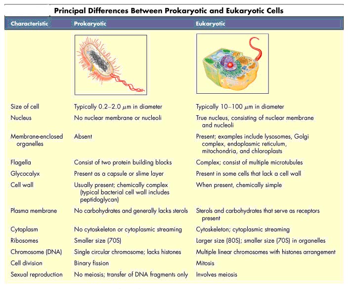
COMPARTMENTALIZATION:
Compaprtmentalization refers to the division of cells into different compartments. Prokaryotes consist of only one compartment containing all the enzymes required for basic functions. Eukaryotes are divided into several compartments. Each compartment has a specific structure and function.
The reasons for the compartmentalization were
a. each compartment performs different functions
b. each compartment has a greater surface area
c. Many enzymes can act closely when it is present in a compartment
d. Cells can become specialized due to the increase in specific compartment to have specific function.
e. Cells become large due to the presence of more compartments.
Unlike in Prokaryotes, the Eukaryotes having different cell organelles to perform different functions. Such as mitochondria is called power house of a cell, ribosomes are called protein factories, lysosomes are called suicide bags, Golgi complexes are called packaging regions etc., ER forms skeletal structure to the cells etc.,
Compartmentalization allows each compartment to perform specific functions without interference from other cell functions. For example, lysosomes can break down cell debris in a compartment without accidentally digesting the cell itself.
It also allows enzymes and substrates to reach higher concentrations than if everything was diluted by the entire cytoplasm. For example, the mitochondria accumulate a large electron gradient in order for the electron transport chain to work.
Origin for Compartmentalization:
The precursors of the first eucaryotic cells are thought to have been simple organisms that resembled bacteria, which generally have a plasma membrane but no internal membranes. The plasma membrane in such cells therefore provides all membrane-dependent functions, including the pumping of ions, ATP synthesis, protein secretion, and lipid synthesis. Typical present-day eucaryotic cells are 10–30 times larger in linear dimension and 1000–10,000 times greater in volume than a typical bacterium such as E. coli. The profusion of internal membranes can be seen in part as an adaptation to this increase in size: the eucaryotic cell has a much smaller ratio of surface area to volume, and its area of plasma membrane is presumably too small to sustain the many vital functions for which membranes are required. The extensive internal membrane systems of a eucaryotic cell alleviate this imbalance.
The evolution of internal membranes evidently accompanied the specialization of membrane function. Consider, for example, the generation of thylakoid vesicles in chloroplasts. These vesicles form during the development of chloroplasts from proplastids in the green leaves of plants. Proplastids are small precursor organelles that are present in all immature plant cells. They are surrounded by a double membrane and develop according to the needs of the differentiated cells: they develop into chloroplasts in leaf cells, for example, and into organelles that store starch, fat, or pigments in other cell types. In the process of differentiating into chloroplasts, specialized membrane patches form and pinch off from the inner membrane of the proplastid. The vesicles that pinch off form a new specialized compartment, the thylakoid, that harbors all of the chloroplast's photosynthetic machinery. Other compartments in eucaryotic cells may have originated in a conceptually similar way. Pinching off of specialized intracellular membrane structures from the plasma membrane, for example, would create organelles with an interior that is topologically equivalent to the exterior of the cell.
The evolutionary scheme described above groups the intracellular compartments in eucaryotic cells into four distinct families: (1) the nucleus and the cytosol, which communicate through nuclear pore complexes and are thus topologically continuous (although functionally distinct); (2) all organelles that function in the secretory and endocytic pathways—including the ER, Golgi apparatus, endosomes, lysosomes, the numerous classes of transport intermediates such as transport vesicles, and possibly peroxisomes; (3) the mitochondria; and (4) the plastids (in plants only).
Each newly synthesized organelle protein must find its way from a ribosome in the cytosol, where it is made, to the organelle where it functions. It does so by following a specific pathway, guided by signals in its amino acid sequence that function as signal sequences or signal patches. Signal sequences and patches are recognized by complementary sorting receptors that deliver the protein to the appropriate target organelle. Proteins that function in the cytosol do not contain sorting signals and therefore remain there after they are synthesized. During cell division, organelles such as the ER and mitochondria are distributed intact to each daughter cell. These organelles contain information that is required for their construction so that they cannot be made from scratch.
Compartments and its functions:
Many vital biochemical processes take place in or on membrane surfaces. Lipid metabolism, for example, is catalyzed mostly by membrane-bound enzymes, and oxidative phosphorylation and photosynthesis both require a membrane to couple the transport of H+ to the synthesis of ATP. Intracellular membrane systems, however, do more for the cell than just provide increased membrane area: they create enclosed compartments that are separate from the cytosol, thus providing the cell with functionally specialized aqueous spaces. Because the lipid bilayer of organelle membranes is impermeable to most hydrophilic molecules, the membrane of each organelle must contain membrane transport proteins that are responsible for the import and export of specific metabolites. Each organelle membrane must also have a mechanism for importing, and incorporating into the organelle, the specific proteins that make the organelle unique.
The nucleus contains the main genome and is the principal site of DNA and RNA synthesis. The surrounding cytoplasm consists of the cytosol and the cytoplasmic organelles suspended in it. The cytosol, constituting a little more than half the total volume of the cell, is the site of protein synthesis and degradation. It also performs most of the cell's intermediary metabolism—that is, the many reactions by which some small molecules are degraded and others are synthesized to provide the building blocks for macromolecules.
About half the total area of membrane in a eucaryotic cell encloses the labyrinthine spaces of the endoplasmic reticulum (ER). The ER has many ribosomes bound to its cytosolic surface; these are engaged in the synthesis of both soluble and integral membrane proteins, most of which are destined either for secretion to the cell exterior or for other organelles. Proteins are translocated into other organelles only after their synthesis is complete, they are translocated into the ER as they are synthesized. This explains why the ER membrane is unique in having ribosomes attached to it. The ER also produces most of the lipid for the rest of the cell and functions as a store for Ca2+ ions. The ER sends many of its proteins and lipids to the Golgi apparatus. The Golgi apparatus consists of organized stacks of disclike compartments called Golgi cisternae; it receives lipids and proteins from the ER and dispatches them to a variety of destinations, usually covalently modifying them en route.
Mitochondria and (in plants) chloroplasts generate most of the ATP used by cells to drive reactions that require an input of free energy; chloroplasts are a specialized version of plastids, which can also have other functions in plant cells, such as the storage of food or pigment molecules. Lysosomes contain digestive enzymes that degrade defective intracellular organelles, as well as macromolecules and particles taken in from outside the cell by endocytosis. On their way to lysosomes, endocytosed material must first pass through a series of organelles called endosomes. Peroxisomes are small vesicular compartments that contain enzymes utilized in a variety of oxidative reactions.
In general, each membrane-enclosed organelle performs the same set of basic functions in all cell types. But to serve the specialized functions of cells, these organelles will vary in abundance and can have additional properties that differ from cell type to cell type.
On average, the membrane-enclosed compartments together occupy nearly half the volume of a cell and a large amount of intracellular membrane is required to make them all. In liver and pancreatic cells, for example, the endoplasmic reticulum has a total membrane surface area that is, respectively, 25 times and 12 times that of the plasma membrane. In terms of its area and mass, the plasma membrane is only a minor membrane in most eucaryotic cells.
Membrane-enclosed organelles often have characteristic positions in the cytosol. In most cells, for example, the Golgi apparatus is located close to the nucleus, whereas the network of ER tubules extends from the nucleus throughout the entire cytosol. These characteristic distributions depend on interactions of the organelles with the cytoskeleton. The localization of both the ER and the Golgi apparatus, for instance, depends on an intact microtubule array; if the microtubules are experimentally depolymerized with a drug, the Golgi apparatus fragments and disperses throughout the cell, and the ER network collapses toward the cell center.
Studying Cells:
Due to the small size of cells, it is difficult to see cells with naked eye. The small size has made the study of cells difficult until the invention of light microscope. However, animal cells are not only tiny but also translucent and colorless. Techniques for understanding the finer structure of cells weren't developed until the latter part of the19th century, when stains that can provide sufficient contrast became available. However, the magnification provided by light microscopes still couldn't resolve the finest details of the cell. It wasn't until the 1940s, when the electron microscope was developed, that scientists were able to visualize the inside of a cell with sufficient detail to understand the internal complexity of the cell. To elucidate even smaller structures inside of the cell such as proteins, X-ray crystallography is used.
Light Microscope
The light microscope can provide enough magnification to visualize the cell. However, because it uses light to resolve objects, the light microscope cannot be used to probe structural details below 500nm. For the most part, scientists using light microscope can see the major organelles of cells such as the mitochondria and nucleus. To help visualize the internal structure of the cell, different organelles could also be selectively stained with different organic dyes such as Malachite green, Sudan black, and Commassie blue.
Different variations of the light microscope have also been created. Currently, the fluorescent light microscope has been widely used to visualize cells stained with different fluorescent dyes. Proteins could also be tagged with fluorescent proteins and visualized under a fluorescent microscope.
Electron Microscope
To increase the resolution of intcs, the electron microscope is developed to use electrons instead of light as the resolving agent. Electrons have a much shorter wavelength than light and can be used to resolve objects on the scale of 0.1nm. However, in order to use the transmission electron microscope, specimens must undergo special preparation. Because electron doesn't have very high penetrating power, all samples must be sliced into 50 to 100nm thin sections.
Besides having high resolution than the light microscope, scanning electron microscopes (SEM) can be used to construct three-dimensional intcs. Here the electron beam is reflected from the surface of the specimen and an object is scanned as a series of layers. The data from the layers are then overlaid on top of each other to create a 3D intc. The resolution of most SEM is about 10nm, so they are mostly used for intact cells and small organisms.
An electron microscope is a type of microscope that uses electrons to illuminate a specimen and create an enlarged intc. Electron microscopes have much greater resolving power than light microscopes and can obtain much higher magnifications. Some electron microscopes can magnify specimens up to 2 million times, while the best light microscopes are limited to magnifications of 2000 times. Both electron and light microscopes have resolution limitations, imposed by their wavelength. The greater resolution and magnification of the electron microscope is due to the wavelength of an electron, its de Broglie wavelength, being much smaller than that of a light photon, electromagnetic radiation. There are 4 types Transmission Electron Microscope (TEM), Scanning Electron Microscope (SEM), Reflection Electron Microscope (REM), Scanning Transmission Electron Microscope (STEM). Electron microscopes are expensive to build and maintain, but the capital and running costs of confocal light microscope systems now overlaps with those of basic electron microscopes.
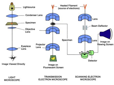
Schematic diagram demonstrating the principal features of a light microscope, a transmission electron microscope, and a scanning electron microscope.
X-Ray Crystallography
X-ray Crystallography is developed to reveal the three-dimensional arrangement of atoms in a molecule. Using x-ray beams with 0.1nm wavelength, a diffraction pattern can be generated by shooting X-ray beams through crystals. The diffraction data can then be used to construct a 3D model of the object. This technique is widely used today to elucidate protein structures.
Reconstructing the intc from the X-ray diffraction data is a tedious and difficult task. Oftentimes, resolution of the structure of one protein requires months of automated data collection and numerous hours of data processing. Sometimes, the slowest step in X-ray crystallography is generation of crystals suitable for producing diffraction patterns.