LYMPHATIC SYSTEM is a system of thin tubes that runs throughout the body. These tubes are called 'lymph vessels or lymphatic vessels’. The lymphatic system is like the blood circulation - the tubes branch through all parts of the body like the arteries and veins that carry blood. Except that the lymphatic system carries a colourless liquid called Lymph.
FUNCTIONS OF LYMPHATIC SYSTEM
The lymphatic system does several jobs in the body. It:-
a. Collects waste materials from tissues.
b. Acts as drainage of metabolites formed in the body.
c. Maintains the body fluids and blood volume.
d. Act as filtering agent and helps in defence mechanism.
e. lymph nodes and other lymphoid tissues are the site of clonal production of immunocompetent lymphocytes and macrophages in the specific immune response.
f. Helps in fat absorption and its transport.
g. Through its reticulo-endothelial system it has phagocytic property.
h. Forms γ-globulins and is concerned with immunologic reactions.
i. Serves to arrest spread of malignant cells although only temporarily.
The lymphatic system may be grouped as:
1.
LYMPHATIC VESSELS:
They resemble veins in structure
except that their coats ae thinner and that these have numerous valves. The
lymph vessels
 are
classified as:
are
classified as:
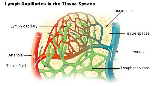
a. Lymph capillaries are situated at sites where lymphatic system begins forming lymphatic plexus. These are larger than blood capillaries. They are formed by endothelial cells that are more elongated in one direction and have connective tissue covering.
b. Medium-sized vessels are formed by union of lymph capillaries. The elongated endothelium is covered by smooth muscle layer of circular and oblique fibres. The connective tissue covering is present above this.
c. Lymphatic ducts are formed by union of medium-sized lymph vessels. They have numerous valves which give them a beaded appearance.
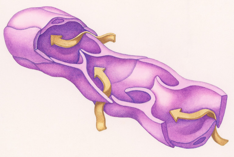
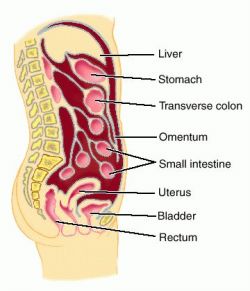
2. SEROUS MEMBRANE: It lines the internal cavity of the body and is reflected over many thoracic and abdominal viscera. The peritoneal serous membrane is the largest in our body. Its inner surface is lined by pavement endothelium, covered by connective tissue.
3.
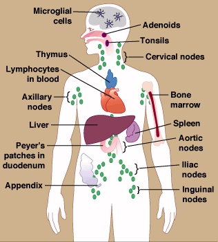
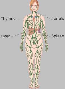
LYMPHATIC TISSUE: It consists of lymph glands, tonsils, thymus, spleen, Payer’s patches, solitary lymph follicles and the appendix.
The lymph vessels develop from various organ systems. In humans, the connection with venous system persists at the opening of thoracic duct and the right lymphatic duct at the entry in to great veins to the root of the neck. Lymphatic system is therefore a closed system.
NERVE SUPPLY TO LYMPHATIC SYSTEM
Lymph vessels are innervated by non-medullated nerves, which end in lymphatic coat by ramification.
LYMPH GLANDS
 Along
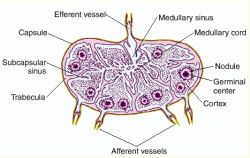
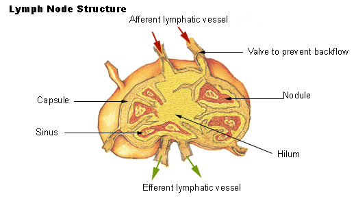
the lymph vessels are small bean-shaped lymph glands or 'nodes'. They are
composed of 2 layers, cortex and medulla, which are lined by
capsular layer of reticular and collagen fibres. These regions are separated all
over by lymph sinuses or channels.
Along
the lymph vessels are small bean-shaped lymph glands or 'nodes'. They are
composed of 2 layers, cortex and medulla, which are lined by
capsular layer of reticular and collagen fibres. These regions are separated all
over by lymph sinuses or channels.
The trabeculae arise inwards from the capsule and after passing through the cortex, these divide and reunite with one another to form a network in the medulla. Hilum is a region formed by depression at one part of the gland and it is the entry point for artery and exit point for veins that supply the glands.
The glandular substance is made up of reticulum. The meshes of reticulum are occupied by lymphoid cells which together form lymph nodules. The nodule has 2 zones:-
1. 1o NODULE: Situated in peripheral cortex. Here, the lymph reaches by afferent lymph vessels which pass through capsule by ramification. The lymphocytes present here are numerous, closed packed and mostly mature in form.
2. 2o NODULE: Also known as germinal centre of Flemming. It is situated in central area of the gland. The lymphoid cells are in lesser number and mostly in precursor form (lymphoblasts). This nodule is the site for lymphocytic development. The cells on maturation get arranged in medullary area in parallel rows and are known as lymph cords. Form these cords, the lymph is carried by efferent lymph vessels which pass out through hilum.
Some of the lymphoid cells in the 2o nodule are converted in to reticulum formation which forms reticulo-endothelial system (RES) in this region.
OTHER LYMPHOID TISSUES
Diffuse Lymphatic Tissue
and Lymphatic nodules:
The alimentary canal, respiratory passages, and genitourinary tract are guarded
by accumulations of lymphatic tissue that are not enclosed by a capsule (i.e.
they are diffuse) and are found in connective tissue beneath the
epithelial mucosa. These cells intercept foreign antigens and then travel to
lymph nodes to undergo differentiation and proliferation. Local concentrations
of lymphocytes in these systems and other areas are called lymphatic nodules.
In general these are single and random but are more concentrated in the GI tract
in the ileum, appendix, cecum, and tonsils. These are collectively called the
GALT
(Gut Associated Lymphatic Tissue).
MALT
(Mucosa Associated Lymphatic Tissue) includes these plus the diffuse lymph
tissue in the respiratory tract.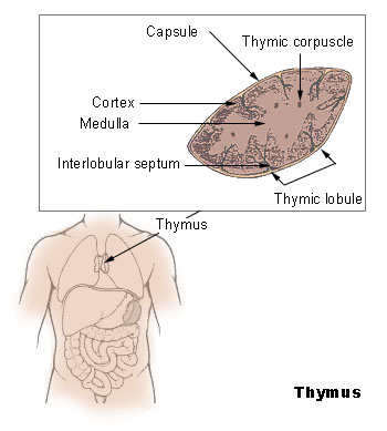
Thymus: The thymus is where immature lymphocytes differentiate into T-lymphocytes. The thymus is fully formed and functional at birth. Characteristic features of thymic structure persist until about puberty, when lymphocyte processing and proliferation are dramatically reduced and eventually eliminated and the thymic tissue is largely replaced by adipose tissue. The lymphocytes released by the thymus are carried to lymph nodes, spleen, and other lymphatic tissue where they form colonies. These colonies form the basis of T-lymphocyte proliferation in the specific immune response. T-lymphocytes survive for long periods and recirculate through lymphatic tissues.
The transformation of primitive or immature lymphocytes into T-lymphocytes and their proliferation in the lymph nodes is promoted by a thymic hormone called thymosin. Ocassionally the thymus persists and may become cancerous after puberty and and the continued secretion of thymosin and the production of abnormal T-cells may contribute to some autoimmune disorders. Conversely, lack of thymosin may also allow inadequate immunologic surveillance and thymosin has been used experimentally to stimulate T-lymphocyte proliferation to fight lymphoma and other cancers.
Spleen: The
spleen filters the blood and rea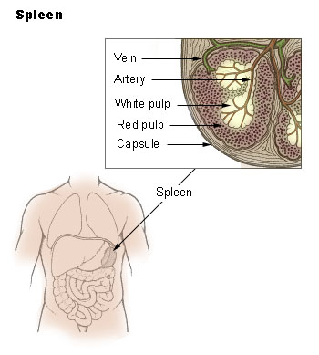 cts immunologically to blood-borne antigens.
This is both a morphologic (physical) and physiologic process. In addition to
large numbers of lymphocytes the spleen contains specialized vascular spaces, a
meshwork of reticular cells and fibers, and a rich supply of macrophages which
monitor the blood. Connective tissue forms a capsule and trabeculae which
contain myofibroblasts, which are contractile. The human spleen holds
relatively little blood compared to other mammals,
but it has the capacity for contraction to release this blood into the
circulation during anoxic stress. White pulp in the spleen contains lymphocytes
and is equivalent to other lymph tissue,
while red pulp contains large numbers of red blood cells that it filters and
degrades.
cts immunologically to blood-borne antigens.
This is both a morphologic (physical) and physiologic process. In addition to
large numbers of lymphocytes the spleen contains specialized vascular spaces, a
meshwork of reticular cells and fibers, and a rich supply of macrophages which
monitor the blood. Connective tissue forms a capsule and trabeculae which
contain myofibroblasts, which are contractile. The human spleen holds
relatively little blood compared to other mammals,
but it has the capacity for contraction to release this blood into the
circulation during anoxic stress. White pulp in the spleen contains lymphocytes
and is equivalent to other lymph tissue,
while red pulp contains large numbers of red blood cells that it filters and
degrades.
The spleen functions in both immune and hematopoietic systems.
a. Immune functions include:
§ proliferation of lymphocytes
§ production of antibodies
§ removal of antigens from the blood
b. Hematopoietic functions include:
§ formation of blood cells during fetal life
§
removal and destruction of aged,
damaged and abnormal red cells and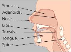 platelets
§ retrieval of iron from hemoglobin degradation
§ storage of red blood cells
Tonsils and Adenoids: These are two glands in the back of the throat. The tonsils and adenoids (also called the 'nasopharyngeal' tonsils) help to protect the entrance to the digestive system and the lungs from bacteria and viruses. The adenoids are at the back of your nose, where it meets the back of your throat.
LYMPH – COLLECTION
F Lymph from various parts of the body is finally collected by 2 main ducts, viz., thoracic duct and the right lymphatic duct, which receive lymph from left and right side of the body, respectively.
F Thoracic duct receives lymph from lower two extremities (legs and fingers), left upper extremity and left side of head, neck and face.
F Right lymphatic duct receives lymph from right side of hand, neck, face, thorax and upper right extremity.
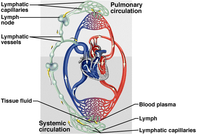
LYMPH – CIRCULATION
In
humans, Lymphokinetic motion
(flow of the lymph) is due to:
¶ Lymph flows down the pressure gradient.
¶ Muscular and respiratory pumps push lymph forward due to function of the semilunar valves.
¶ Lymph flow is brought by contraction of lymph vessels.
¶ Presence of valves makes one-way flow and prevents regurgitation.
¶ Plymph vessels = 4 – 8 mm H2O while, Pthoracic duct = 0 – 6 mm H2O. This difference in pressure gradient contributes to the flow.
¶ Similarly, P tissues - Plymph vessels pressure gradient maintains flow from tissues to lymph vessels.
¶ The suction action of thorax is also an additive factor, which is aided by diaphragm during respiration.
v The most important function of lymphatic circulation is removal of proteins from interstitial spaces. These proteins and other large size particulates cannot be removed through the blood capillaries.
RATE OF LYMPH FLOW: 1 – 1.5 mL/min. Of the 120 mL/h, 100 mL passes through thoracic duct and 20 mL through other channels.
Rate of lymph flow = Ptissue x Activity of lymphatic pump
FACTORS AFFECTING RATE OF LYMPH FLOW ARE:
a) ^ Pcapillary ; b) ^ Capillary permeability; c) ^ tissue fluid proteins; d) Decrease in plasma Poncotic
LYMPH – COMPOSITION (RETICULO-ENDOTHELIAL SYSTEM)
|
SUBSTANCES |
AMOUNT |
|
H2O |
94% |
|
Proteins |
2 – 4.5 g% |
|
Carbohydrates |
130 mg% |
|
Urea |
20 – 25 mg% |
|
NPN |
34 mg% |
|
Creatine |
1.5 mg% |
|
Chlorides |
700 mg% |
|
Ca2+ |
9 – 10 mg% |
|
Pi |
5.5 mg% |
|
Fat |
Low during fasting, but ^ ses to 5 – 15% after meals |